• English
  • Ziehm Member Service
  • Ziehm Academy
  • Products
  • Business Success
  • Clinical Evidence
  • Contact Us
English
  • Home
  • Products
    • C-arms with Flat-Panel Detector
      • Ziehm Vision RFD 3D
      • Ziehm Vision RFD Hybrid Edition
      • Ziehm Vision RFD
      • Ziehm Vision FD
      • Ziehm Solo FD
    • C-arms with 3D Imaging
      • Ziehm Vision RFD 3D
    • Orthoscan Mini C-arms
      • Orthoscan TAU 2020
      • Orthoscan TAU 1512
      • Orthoscan TAU 1515
      • Orthoscan Mobile DI
  • Business Success
    • Innovation and Technology Leader
    • Investment Pays Off
  • Clinical Evidence
    • Focus Topic Precision
    • Ease of Use
    • Image Quality
    • The Right Dose
    • Clinical Applications
  • About Us
    • Company
    • Contact Us
    • Daughter Companies
    • Cooperation Partners
    • Locations
    • Managing Directors
    • Sustainability
  • Career
    • Working at Ziehm Imaging
  • Global Service
    • Ziehm Tailored Solutions
    • Service Locations
Ziehm Member Service Ziehm Academy Facebook Youtube Linkedin Instagram

  • Ziehm Imaging
  • Clinical Evidence

Image Quality CHANGING THE GAME

Image Quality

The core of medical imaging: Image quality

In medical imaging, the most important and desired goal is to benefit from the best clinical image quality. Being the innovation leader on the market, Ziehm Imaging provides a range of hard- and software features in their mobile C-arms, offering superior image quality. This changes the game for our clinicians and let them discover new areas of clinical applications.

Image Quality

High-end vascular surgery

This final angiogram shows the successful implantation of three branches in a fenestrated EVAR. More than 65,000 grayscales taken by Ziehm Imaging's flat-panel detector help to visualize the exact trace and allows a much easier differentiated assessment of the vessels.

Courtesy
Hospital Egas Moniz, Lisbon, Portugal

Device used
Ziehm Vision RFD Hybrid Edition1

Image Quality

Unmatched mobile 3D imaging

Visualizing even the smallest anatomical details is one of the most challenging parts in mobile 3D imaging. In this case, we show the finest anatomical details in maxillofacial surgery in razor-sharp quality. With an intraoperative control scan, unnecessary revisions can potentially be reduced and the radiation dose of postoperative CT scans avoided.

Courtesy
Berufsgenossenschaftliche Unfallklinik Murnau (Murnau Trauma Center), Germany

Device used
Ziehm Vision RFD 3D

Image Quality

Image of the year 2017

This case shows the sophisticated endoprosthetic surgical repair of an abdominal aortic aneurysm performed using an innovative endovascular sealing technology (EVAS). Here, the Advanced Active Cooling technology significantly increases the performance capacity by avoiding overheating of the C-arm. ZAIP (Ziehm Adaptive Image Processing) ensures crystal-clear resolution of the stent. The noise suppression in the background also helps create outstanding image quality.

Courtesy
University Hospital Parma, Italy

Device used
Ziehm Vision RFD Hybrid Edition1


Curious to discover more?

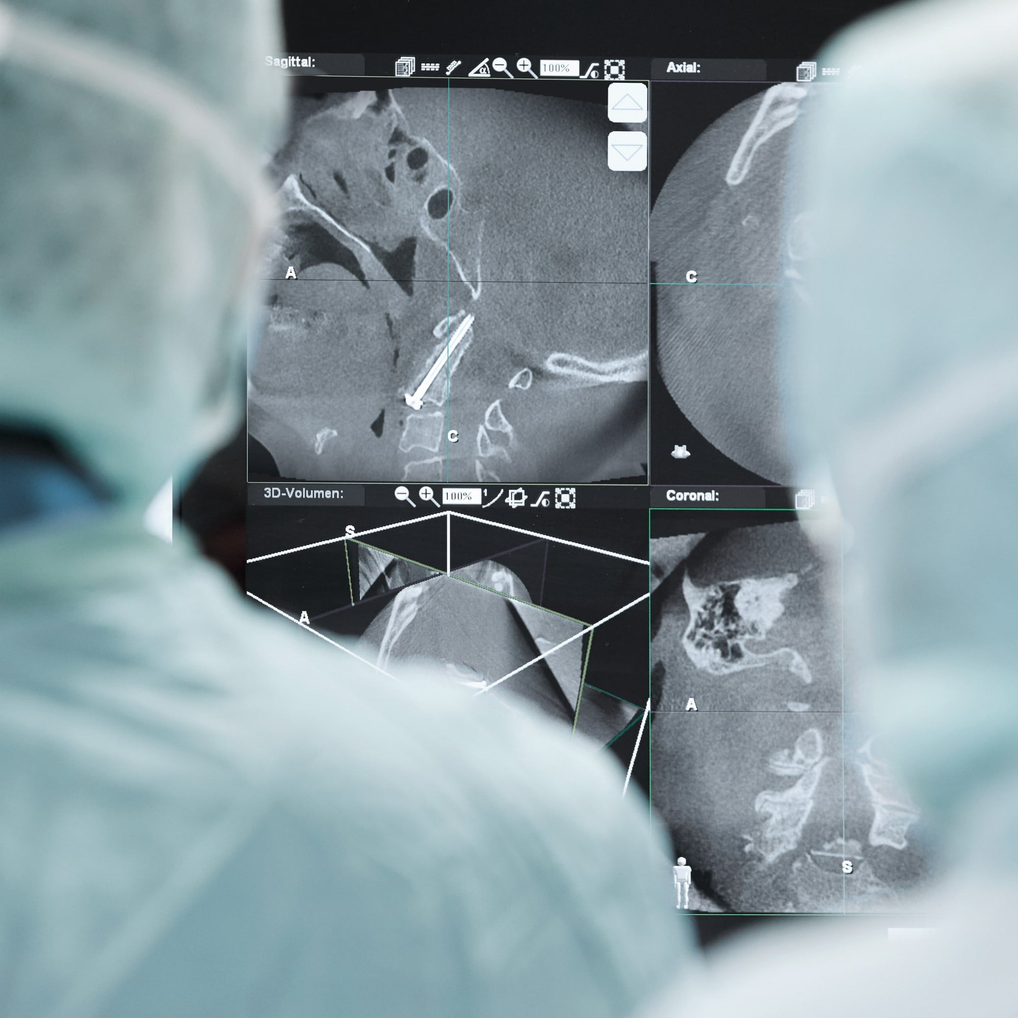
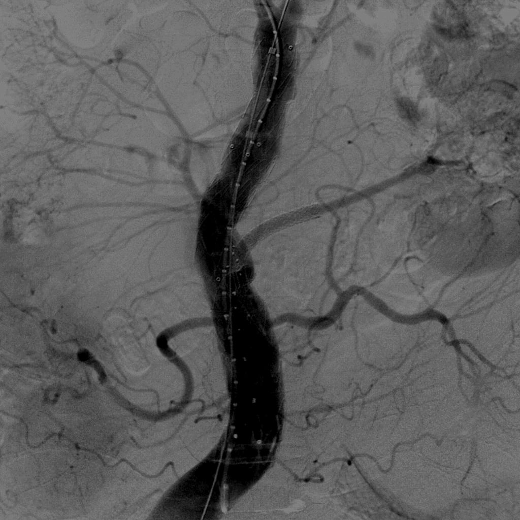
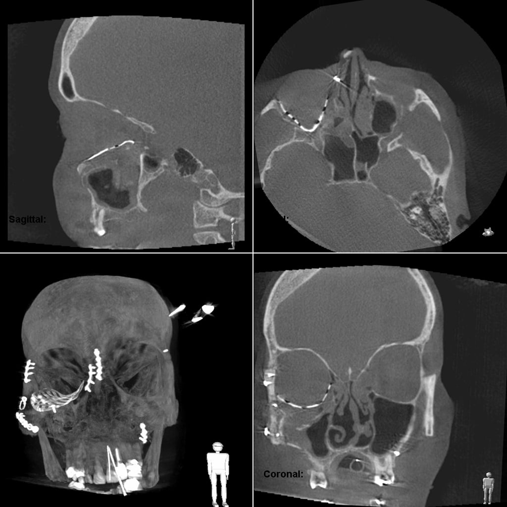
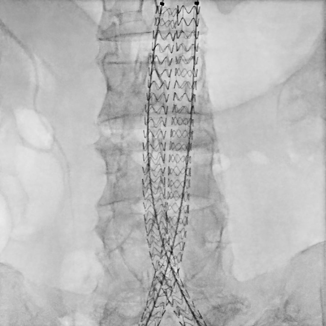
Find a selection of the best clinical images in our interactive image gallery

Ziehm image gallery

1The Ziehm Vision RFD Hybrid Edition represents a group of optional hardware and software that creates an option package on the device named Ziehm Vision RFD.

Tailored Solutions

  • Products
  • Clinical Evidence
  • Business Success

Support

  • Global Service
  • Ziehm Member Service
  • Ziehm Wiki
  • Ziehm Academy
  • Downloads

About

  • News
  • Events

Follow us

  • Facebook
  • Youtube
  • Linkedin
  • Instagram
  • Legal Notice
  • Site Owner
  • Privacy
Copyright © 2023 Ziehm Imaging GmbH