
Library
Clinical Stories

Navigating the change
Panorama
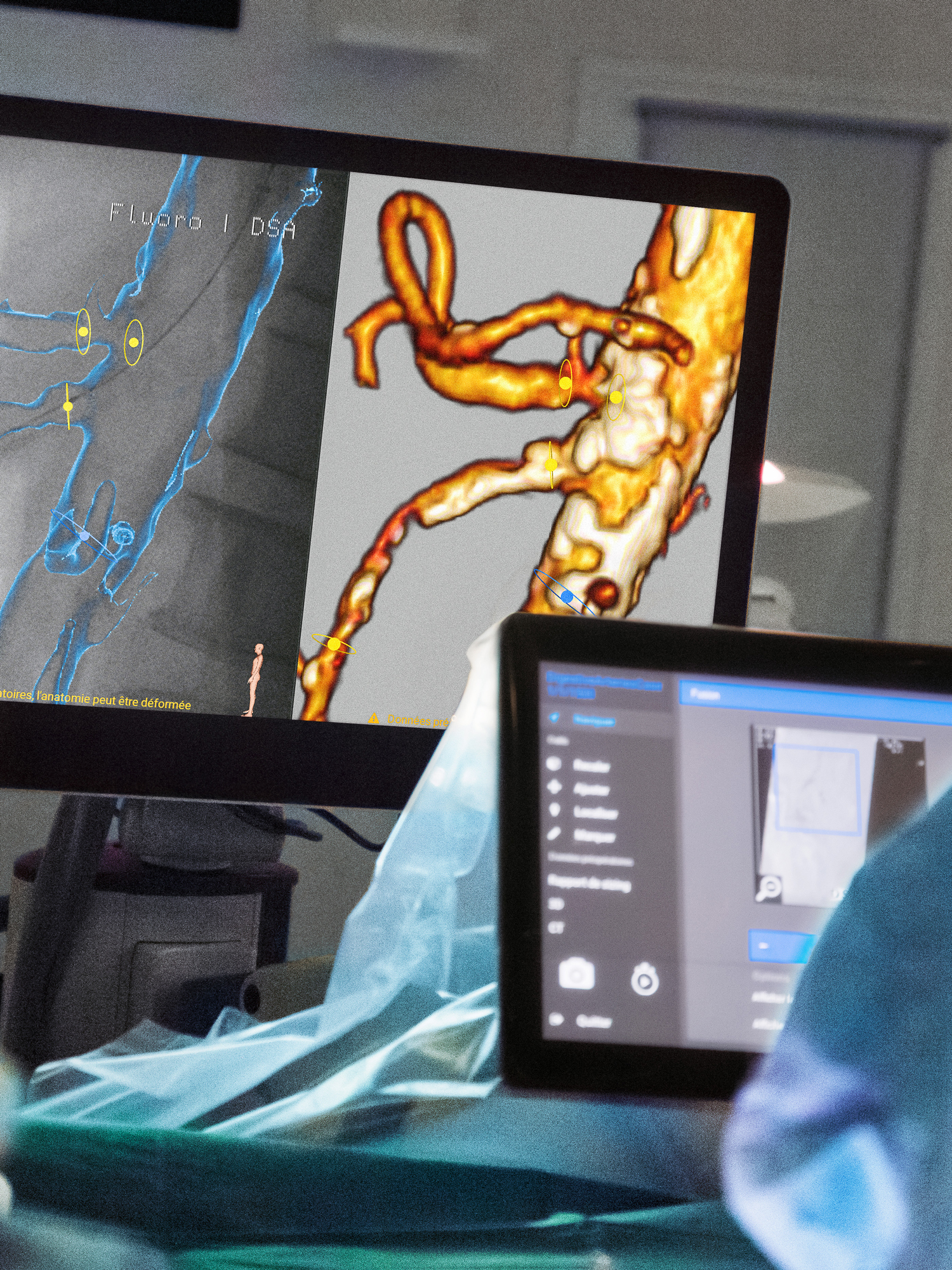
Fusion
Learning from India
Certainty in real-time
Before the first cut
With precision to perfection
The highest level of care
The future is mobile
Interviews
Synergies
Operating the system
Mr. C(EO)MOS
Features
Three values
Tips & Tricks
Case planning in minutes
The mobile CathLab
Three steps to the perfect vascular image
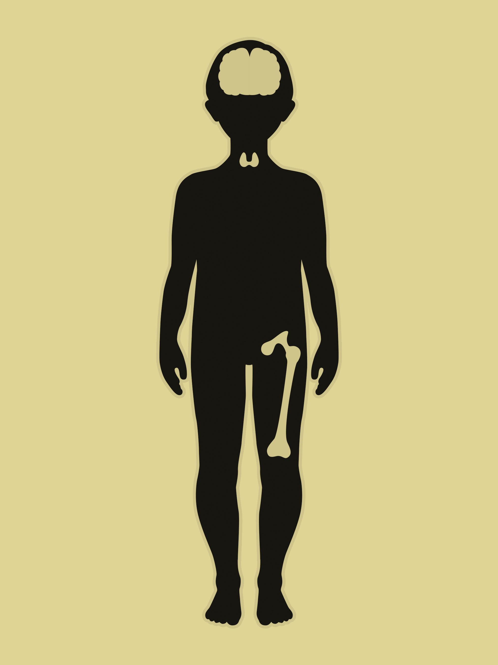
Radiation protection in pediatrics
Three steps for creating a 3D image
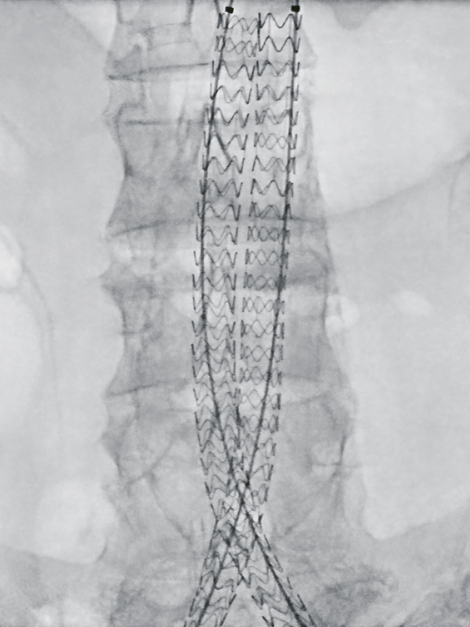
Image of the Year
2022

2020
2019
2018
2017
People
Prof. Dr. Diana Arweiler-Harbeck: Three values, Feature
Prof. Dr. Wolfgang Böcker: The highest level of care, Clinical Story
Prof. Dr. Wolfgang Böcker: Three values, Feature
Dr. Ellen Frydenberg: With precision to perfection, Clinical Story
Heidi Garberg: Before the first cut, Clinical Story
Cemil Göksu: Fusion, Clinical Story
Bernhard Hochholdinger, Ing. Mag. (FH): Operating the system, Clinical Story
Jorunn Hommelstad: Before the first cut, Clinical Story
Klaus Hörndler: Mr. C(EO)MOS, Interview
Prof. Dr. Christoph Josten: Synergies, Interview
Prof. Dr. Christoph Josten: Three values, Feature
Prof. Dr. Adrien Kaladji: Fusion, Clinical Story
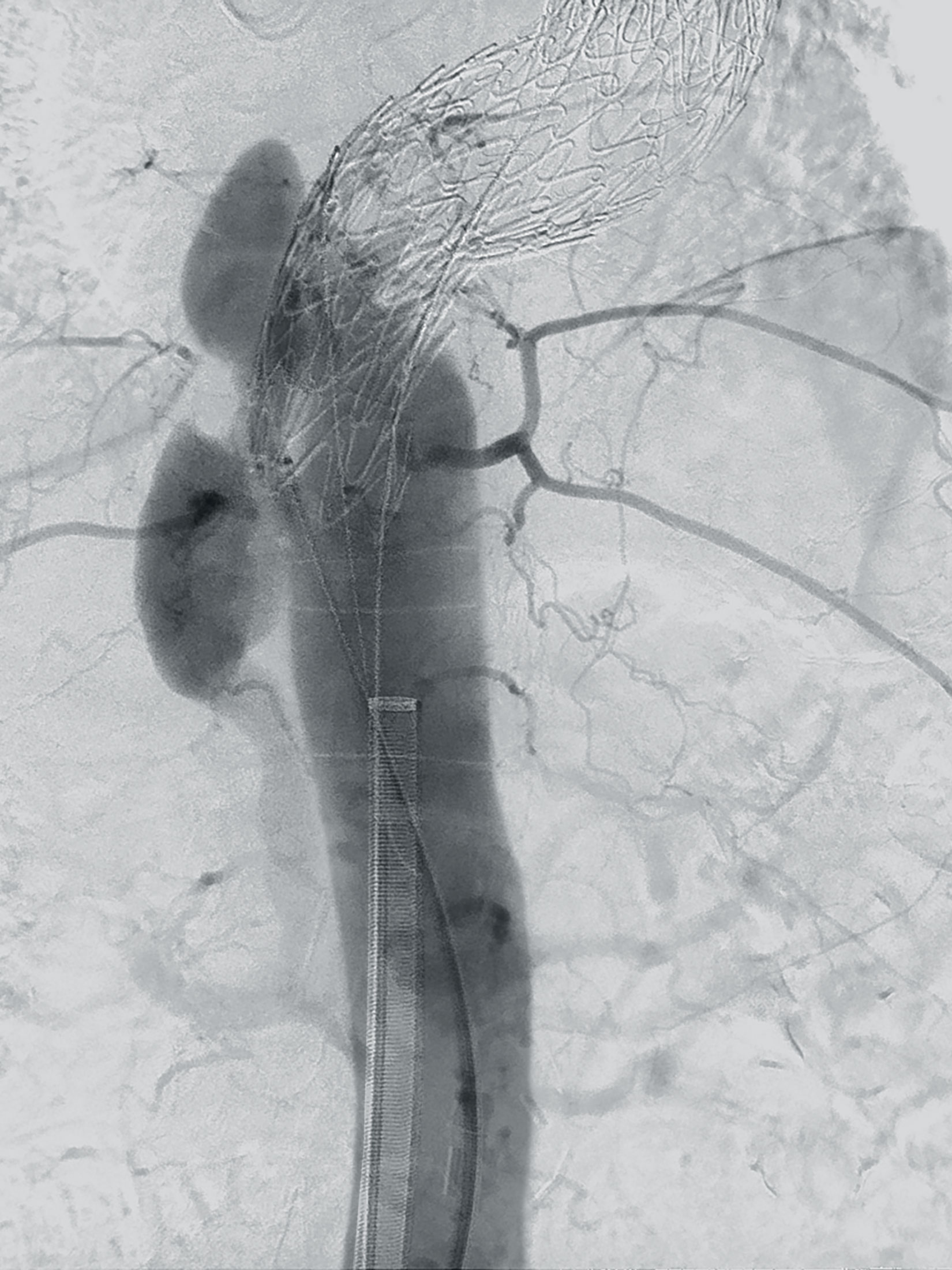
Prof. Dr. Johannes Kalder: Panorama, Clinical Story
Priv.-Doz. Dr. Andreas Kammerlander: The highest level of care, Clinical Story
Priv.-Doz. Dr. Paula Keschenau: Panorama, Clinical Story
Norbert Lechner, Ing.: Operating the system, Clinical Story
Anne Lindemann: Panorama, Clinical Story
Dr. Aimo Miettinen: Image of the Year 2022
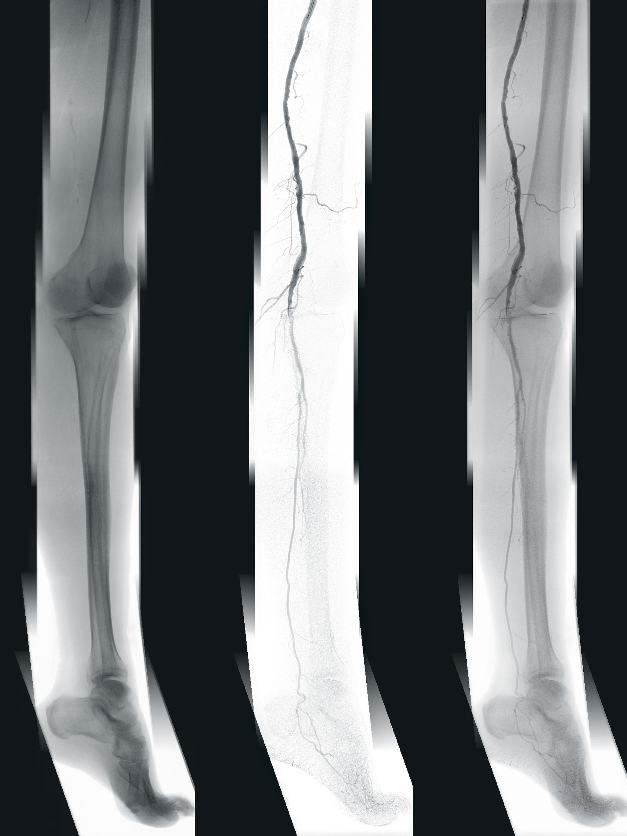
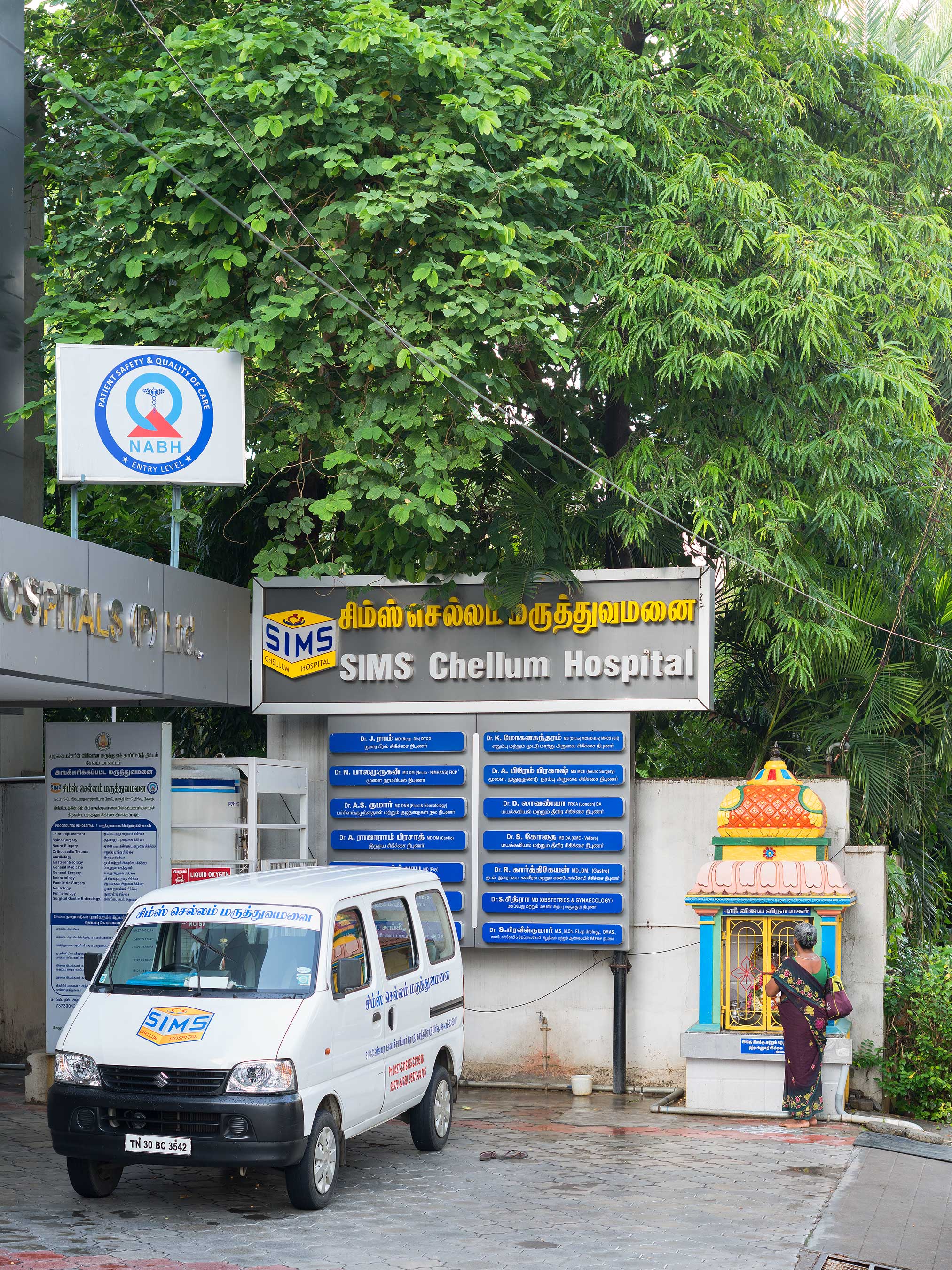
Dr. Rajaram Prasad: Learning from India, Clinical Story
Trude Rosvold: Before the first cut, Clinical Story
Dr. Milton Routt: Three values, Feature
Dr. Sven Seifert: The future is mobile, Clinical Story
Dr. Timothy Steel: With precision to perfection, Clinical Story
Prof. Dr. Gian Franco Veraldi: Certainty in real-time, Clinical Story
Dr. Simon Weidert: The highest level of care, Clinical Story
Priv.-Doz. Dr. Christian Zeckey: The highest level of care, Clinical Story