The highest
level of care
Photos
Juli Sing
With over thirty operating rooms, the Operating Center in Munich at the Grosshadern campus is one of the largest in Europe. In the Department for General, Trauma and Reconstructive Surgery under the direction of Prof. Dr. Böcker, physicians use new methods and the most advanced medical technology to provide acute treatment for many injured patients.
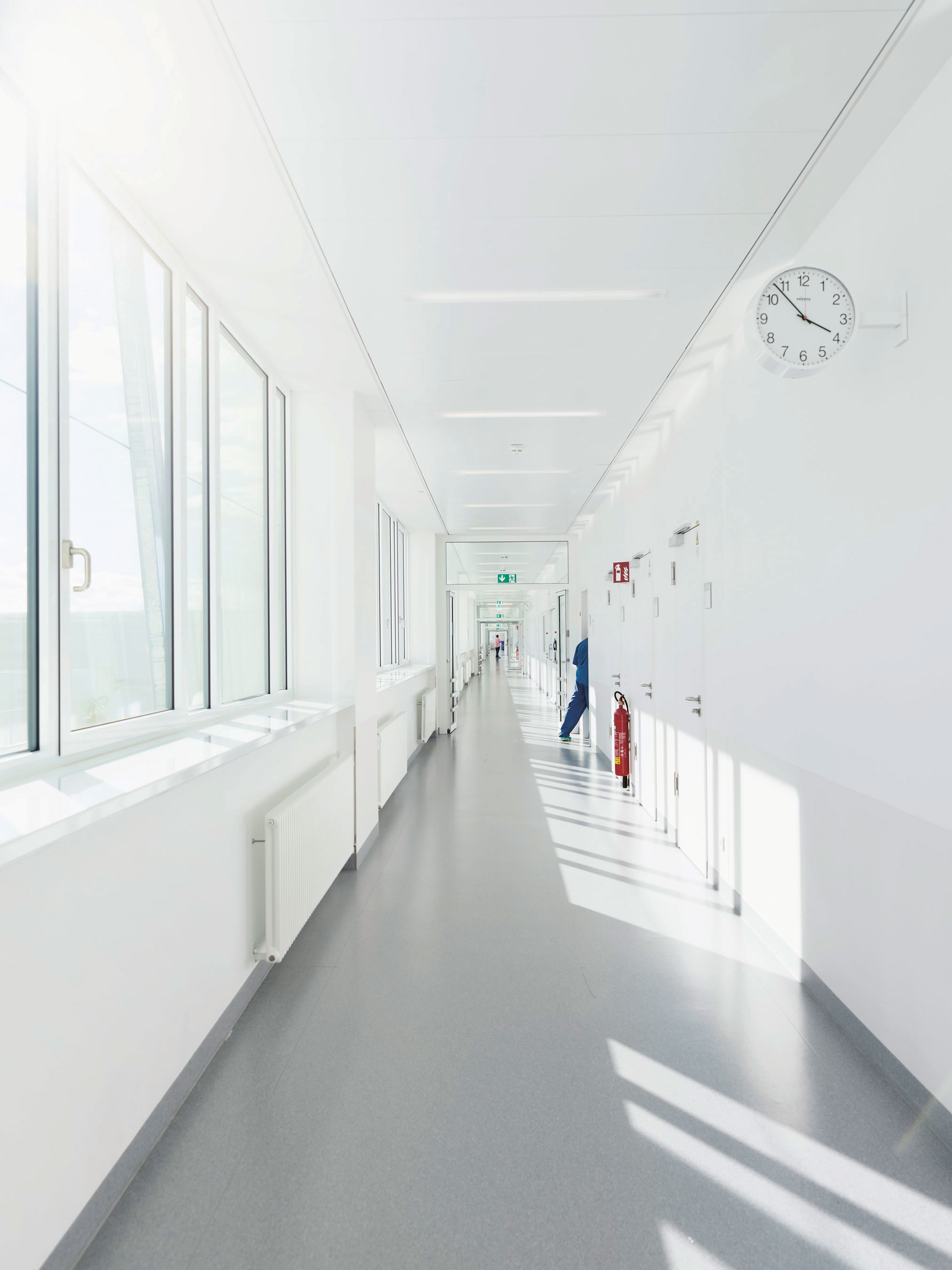
The long halls of the Operating Center in Großhadern provide a sense of the size of this large building. More than 40,000 operations are performed each year here since the center opened in 2014. The Operating Center, part of the clinic at Ludwig-Maximilians-University (LMU), is considered a successful model in professional circles. The influence of the LMU is clear: advanced, minimally invasive procedures, operating in hybrid rooms, and using a sliding gantry, i.e. computed tomography equipment that can be moved back and forth between two operating rooms demonstrate the close connection to research.
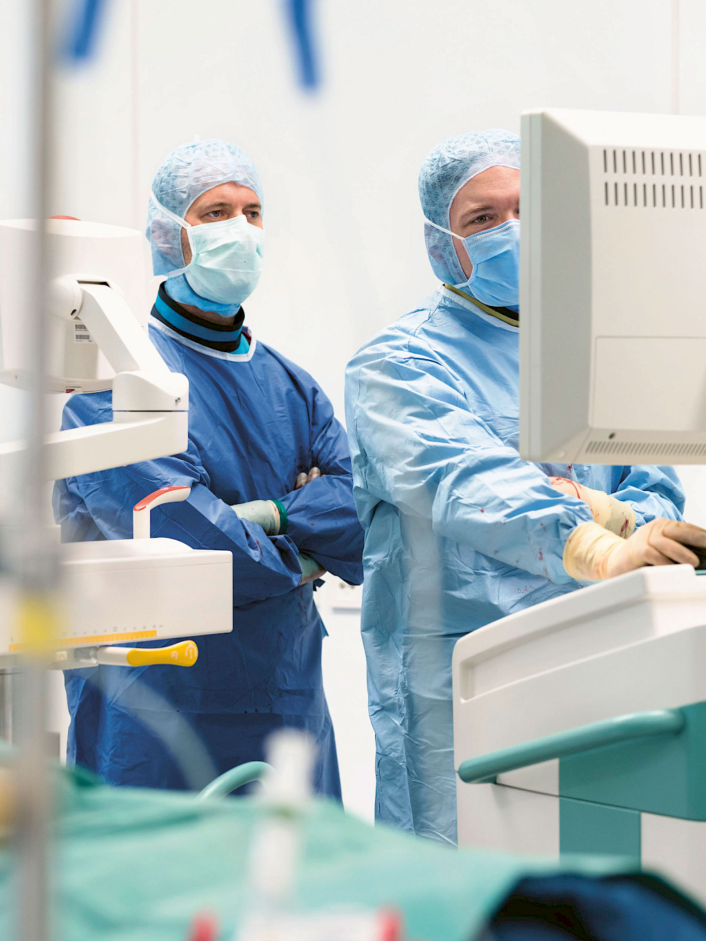
But the standard technology is highly advanced as well. From patient entrance areas and procedures to the central sterile material supplies department and on to intraoperative imaging, medical advances are a great priority everywhere in the Operation Center. For this reason, in the spinal and pelvic surgery section of the Department for General, Trauma and Reconstructive Surgery, senior physician Dr. Zeckey and specialist Dr. Weidert are planning today’s procedures with the assistance of the newest imaging systems.
Both doctors work on the team of Dr. Kammerlander, the head physician. He and Prof. Dr. Böcker, the clinic director, are known internationally as experts in their field. That’s why, again and again, patients from all over Europe are flown to Großhadern to undergo a second procedure performed by professionals. Prof. Dr. Böcker’s team includes nearly fifty doctors, more than twenty of whom are head physicians or specialists. The clinic, a verified mass casualty trauma center, offers treatment for a wide range of situations: from broken bones and tissue damage to athletic injuries and severely injured patients.
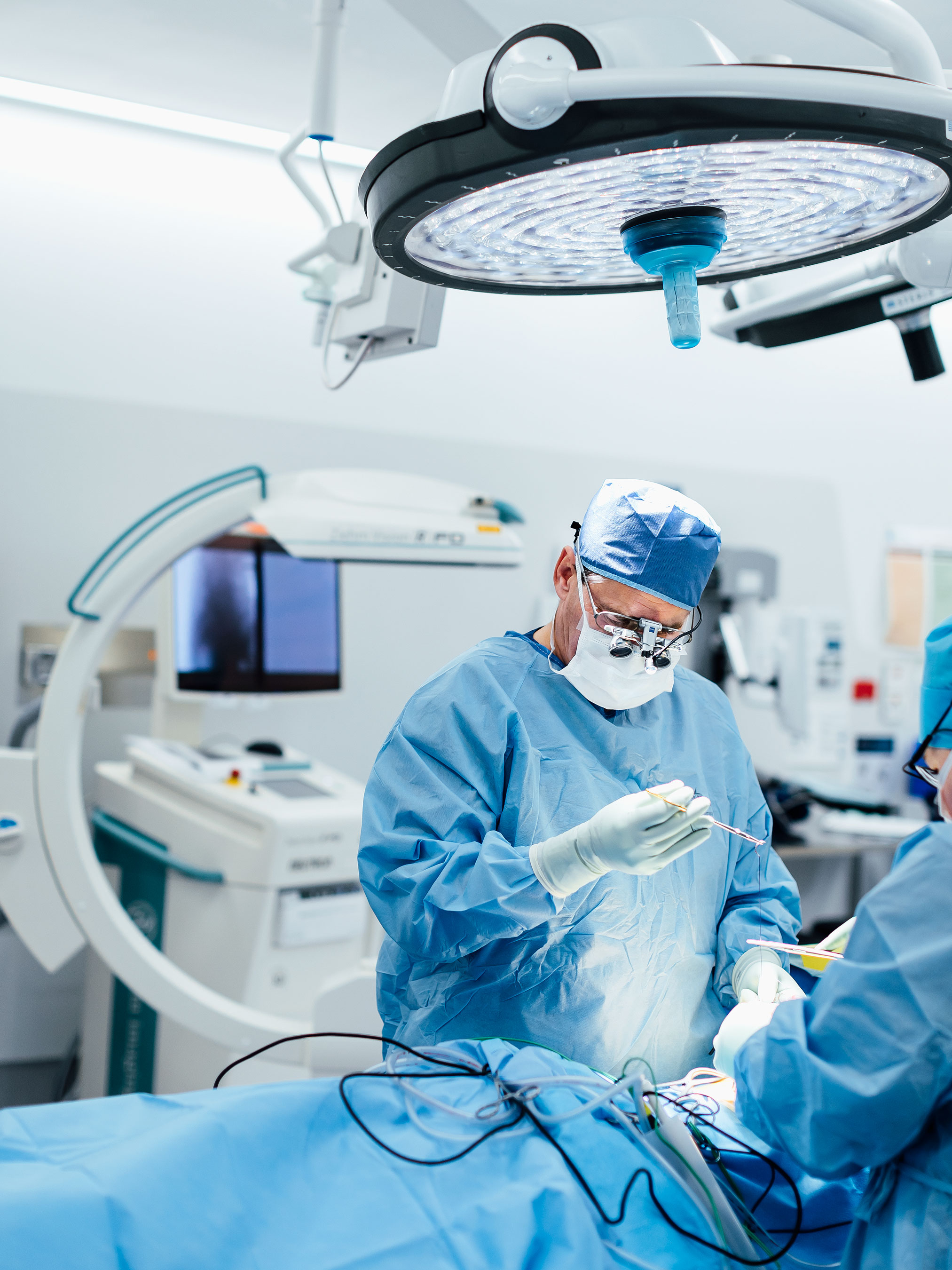
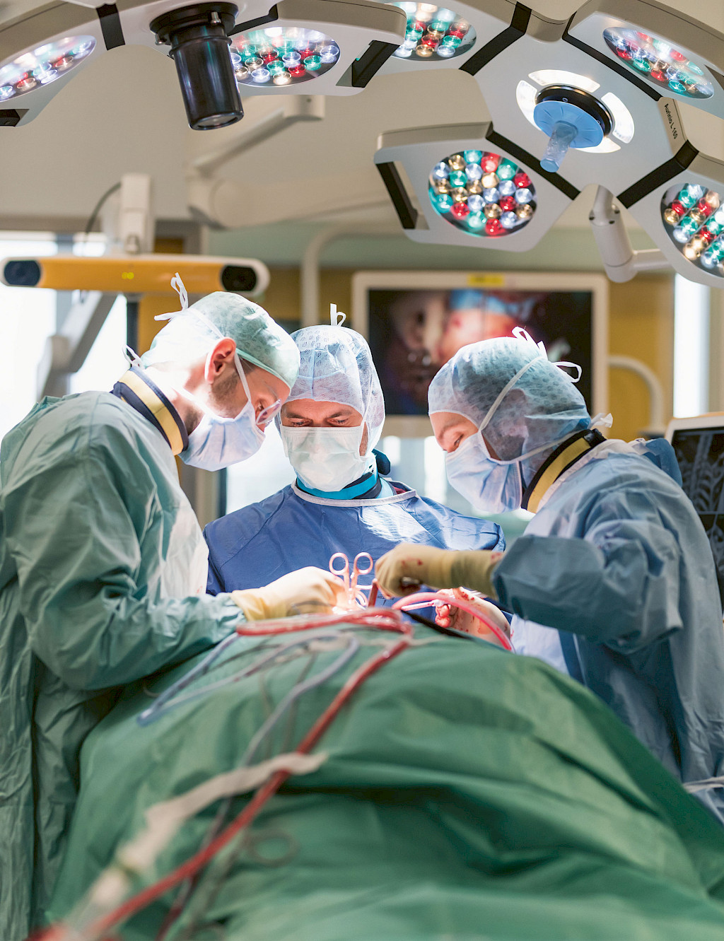
Today Dr. Zeckey and Dr. Weidert are performing two spinal procedures. The first operation on the cervical spine is especially challenging: At the patient’s request, the surgeons are using an alternative method of treatment. Dr. Zeckey and Dr. Weidert discuss the procedure in detail: because each action must be correct, advanced medical technology is used even in the planning phase.
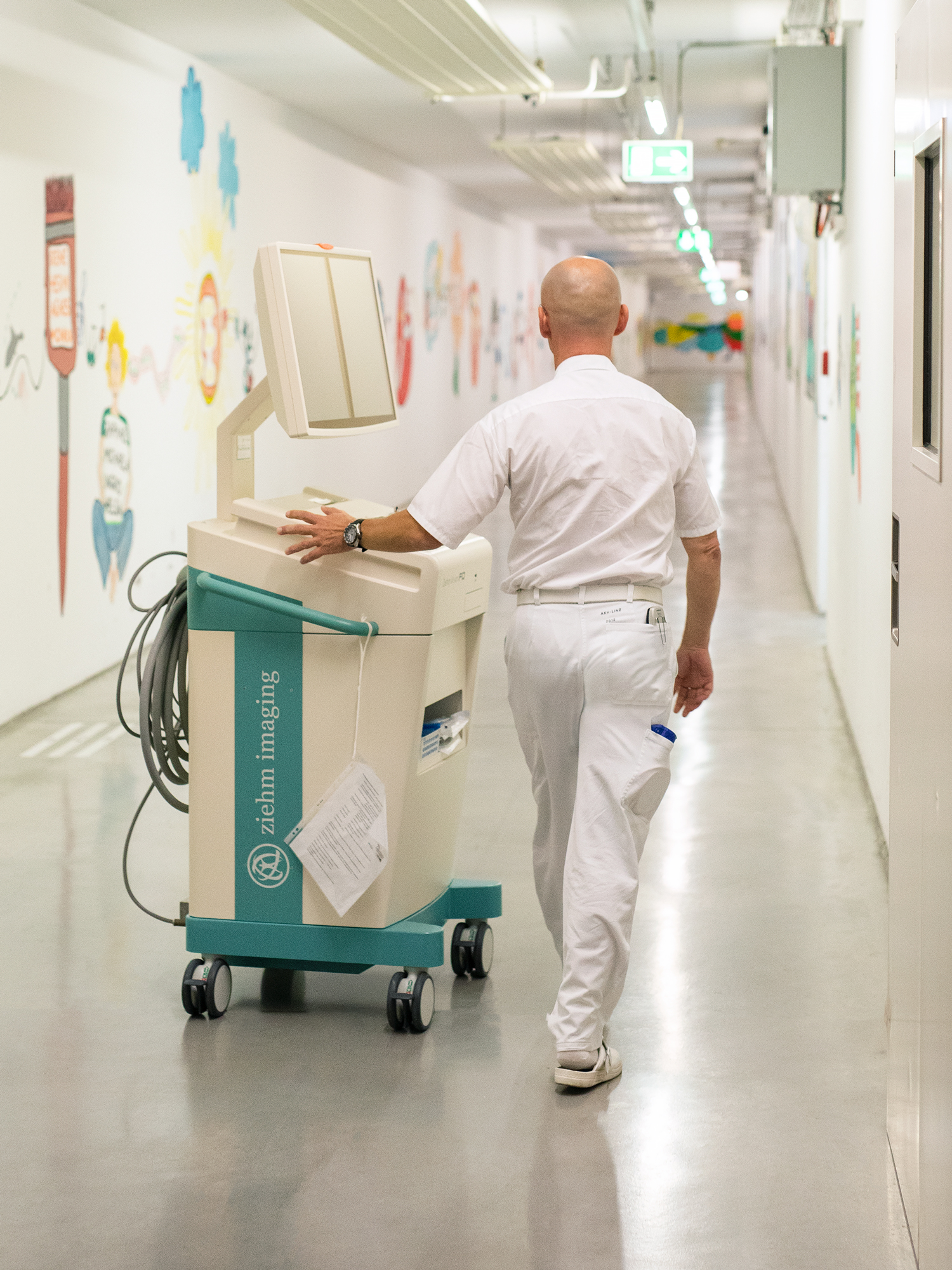
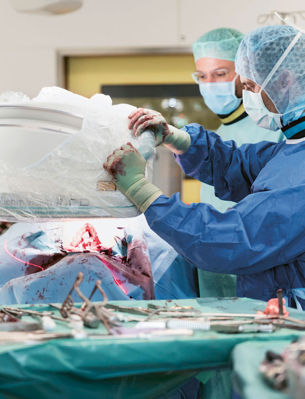
For today's procedure, the physicians are using a new 3D C‑arm by Ziehm Imaging: the Ziehm Vision RFD 3D with premium CMOSline1 equipment, which will be introduced to the market in just a few months. As a reference partner, the trauma surgery department receives the system as a loan unit much earlier in order to test the technical innovations in clinical use. A new detector, settings that reduce radiation exposure, and expanded user functions have improved the image quality of the 3D C‑arm. Today the new system has to demonstrate its capabilities.
Operating on the cervicothoracic area, the junction between the cervical spine and the thoracic spine, is difficult enough, and this, together with the anatomical condition of the patient, makes a precise lateral 2D aquisition of the transition to the thoracic spine nearly impossible. But the high contrast image of the bony structures is an important requirement for the success of the operation. If the vertebral bodies cannot be represented precisely with intraoperative imaging, the spinal cord could be damaged, for example, due to the improper placement of screws. That’s why Dr. Zeckey and Dr. Weidert have decided to perform the operation using image-guided navigation and to scan the spine with the mobile 3D C‑arm.
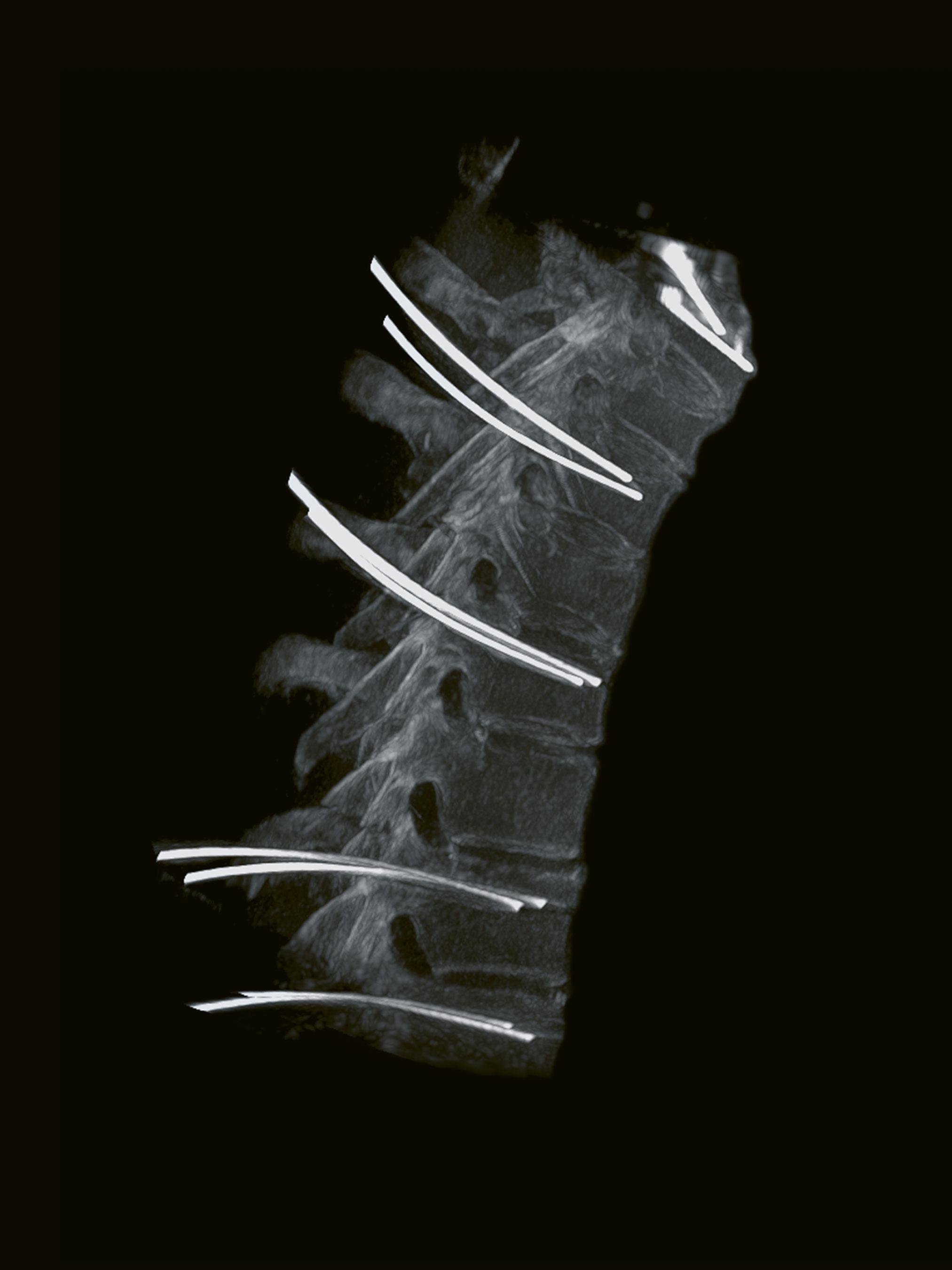
The C‑arm moves 180° around the patient in both linear and rotational motion. The 3D data set aquired in this way displays even the smallest anatomical details of the vertebral bodies. The physicians use this as a starting point for image-guided navigation during the operation. First, based on this 3D data set, they plan the exact placement of the screws on the monitor, and then later, during the operation, they display the progress made in setting the screws.
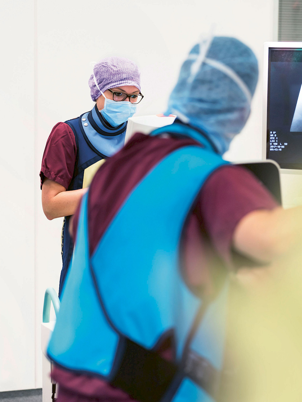
Application Specialist Nadja Baitis operates the C‑arm. She is a trained medicaltechnical radiology assistant and is part of 3D product management at Ziehm Imaging. In particular, Ms. Baitis works with new C‑arm systems during the test phase to ensure that all of the technical innovations also function in practical use. She trains the OR personnel with regard to using the system and passes on her valuable experience. Ms. Baitis has been in Großhadern for more than two weeks already and is testing the new system in clinical applications. For difficult procedures like the one today, she controls the C‑arm herself. The operation on the cervical spine takes more than four hours and the Ziehm Vision RFD 3D CMOSline is consulted for clinical guidance again and again. Finally, the team carries out a 3D scan.
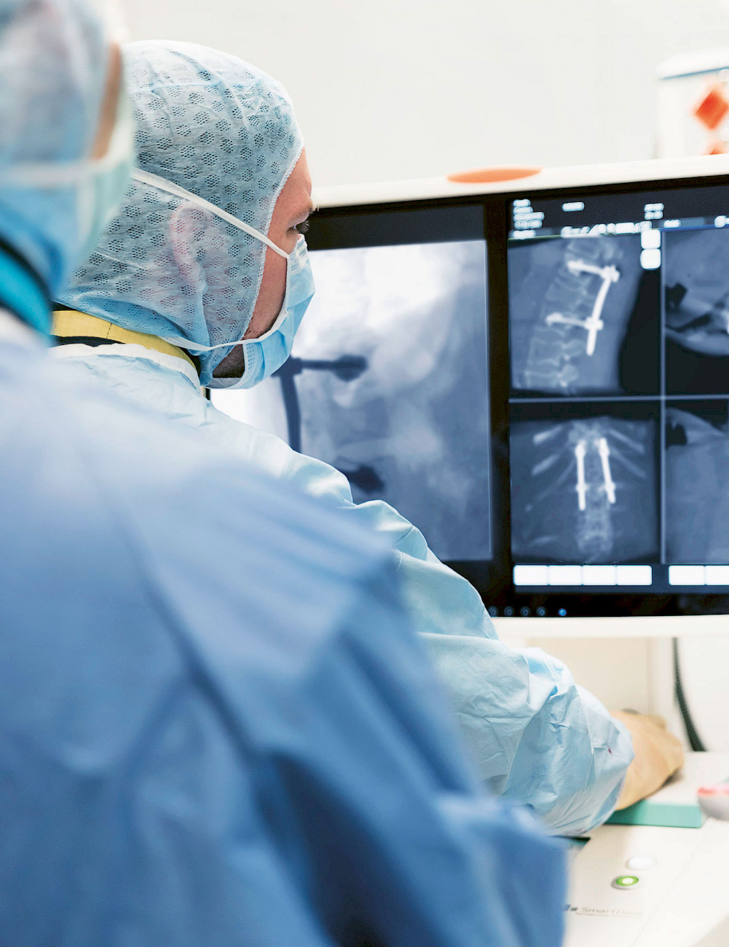
Because the implant can be positioned precisely and this intraoperative control scan can be performed, the patient does not have to wait for the results of a post-operative CT scan and perhaps undergo another operation. The final 3D image confirms it: The screws are correctly positioned in the bone at the specified anatomical distance from the arteries and spinal cord. The operation was successful. The Ziehm Vision RFD 3D CMOSline is also used in the next operation, a dorsal instrumentation procedure in the lumbar spine.
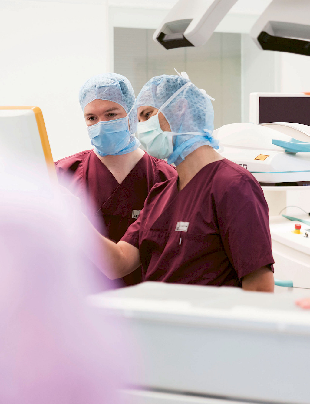
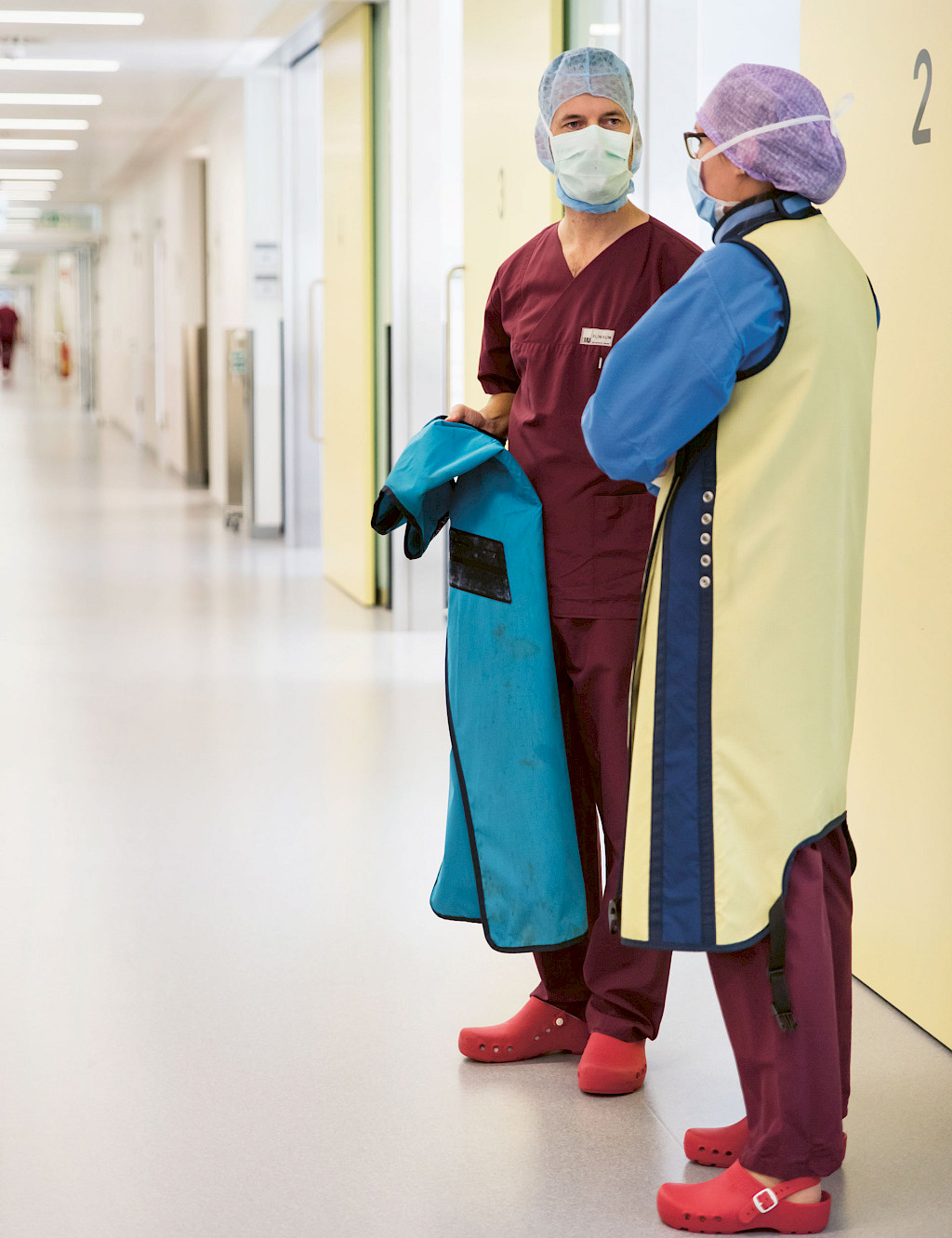
Following training by application specialist Ms. Baitis, Dr. Weidert operates the 3D C‑arm himself from the sterile area. Once again, the intraoperative 3D imaging provides assurance that the implants are placed exactly where they should be to prevent pain and alleviate the need for further treatment. Following the procedure, Ms. Baitis consults with the physicians.
How does the team rate the device’s performance? Senior physician Dr. Zeckey is impressed by the quality of the Ziehm Vision RFD 3D CMOSline. And head physician and deputy clinic director Dr. Kammerlander is also satisfied. He uses intraoperative navigation regularly in his standard operation practice. The high-resolution imaging together with the navigation provides assistance, particularly in anatomical areas that are difficult to see into, such as the cervicothoracic transition. The image-guided navigation function allows him to achieve the best possible results in spinal and pelvic procedures.
The director of the Department for General, Trauma and Reconstructive Surgery, Prof. Dr. Böcker, is also very pleased with the larger volume size of the 3D C‑arm. This new option provides a larger scanning area, which allows more anatomy to be displayed in multiplanar reconstruction. For a large anatomical region such as the pelvis, a single 3D scan now provides adequate coverage of the complete operating area. Ms. Baitis values the close cooperation with the trauma surgeons. She trusts the expertise of the physicians in Großhadern and is glad that the CMOSline version of the Ziehm Vision RFD 3D passed the first practical test. She will be here at the Operating Center in Großhadern again tomorrow for the C‑arm’s next application.
Disclaimer
1
The CMOSline is a system configuration based on the Ziehm Imaging CMOS flat-panel detector.
More information on Ziehm Vision RFD 3D
—
This clinical story was published in issue 2.
Download issue 2 as PDF