Three values
Illustrations
Sebastian Mildenberger
In three-dimensional space, three values are required to determine the position of an element. Within three coordinate axes, we can create a reconstruction of the space inside the human body. And in recent years, such 3D images have revolutionized operating rooms. Highly valued by surgeons, these images contain more than axial, coronal and sagittal views of the body. Clinical images from the physicians‘ everyday practice show us the benefits of this technology.
Spine and pelvis surgery
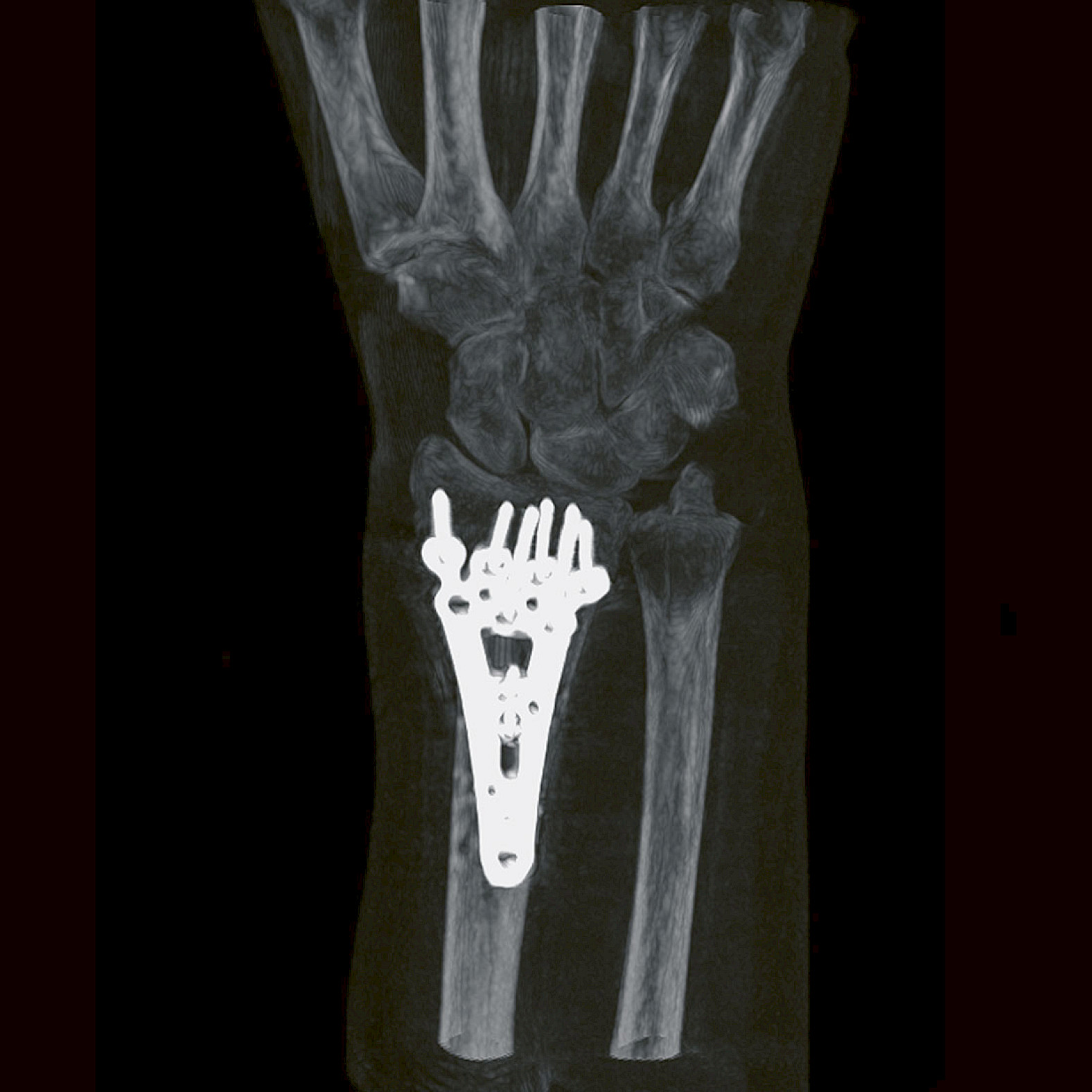
“Intraoperative 3D imaging is recognized as the most advanced mobile device imaging technology for achieving complete CT-like image information in just one scan procedure. For us, the ability to switch seamlessly among intraoperative navigation, CT-like 3D imaging, and 2D fluoroscopy provides an ideal combination for daily spine and trauma cases such as this particular distal radius fracture.”
— Professor Wolfgang Böcker
University Hospital Munich, Germany
Medical product design has its own set of rules. It’s about more than aesthetically pleasing shapes or haptics. Healthcare technology must meet high requirements for safety and hygiene, reflect state-of-the-art research, and feature handling that is clearly understandable and intuitive. A look at the design information for the Ziehm Vision RFD 3D demonstrates the wide variety of dimensions involved in planning all of the details.
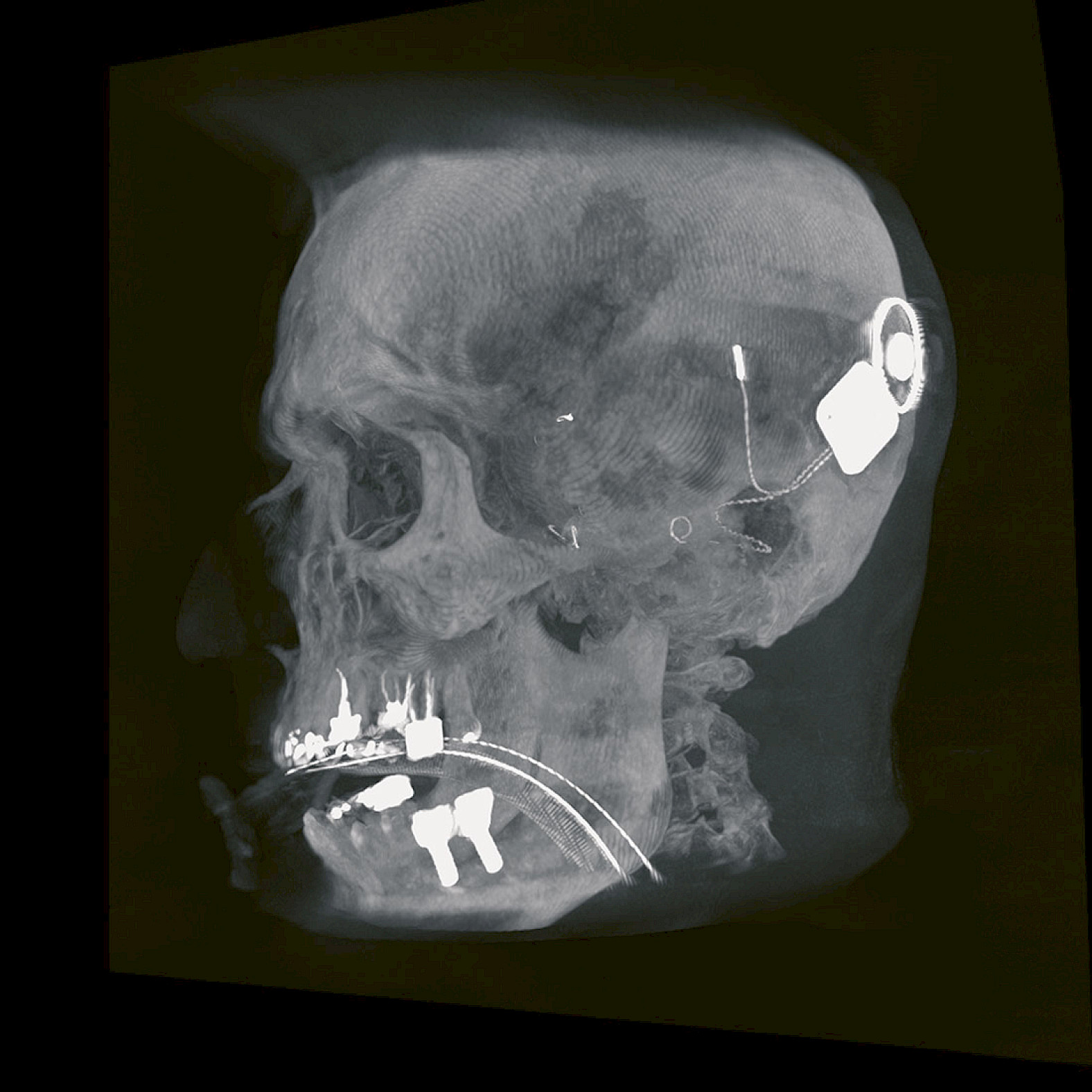
Cochlear implant
“The cochlear implant is an inner ear prosthesis that rehabilitates severe to profound sensorineural hearing loss in patients who haven’t benefitted from conventional sound amplification. 3D imaging plays a vital role for us in planning, implementation, control and, therefore, the successful placement of a cochlear implant. Especially the new, higher resolution of 512 voxels significantly improves the visualization of details, with razor-sharp images of even the smallest anatomical structures in the middle and inner ear as well as the tiny electrode contacts within the cochlea. The result is a highly successful hearing outcome, providing our patients with great improvements in their everyday lives.”
— Professor Dr. Diana Arweiler-Harbeck
University Hospital Essen, Germany
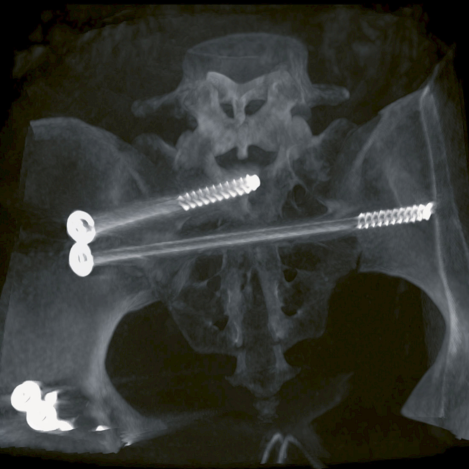
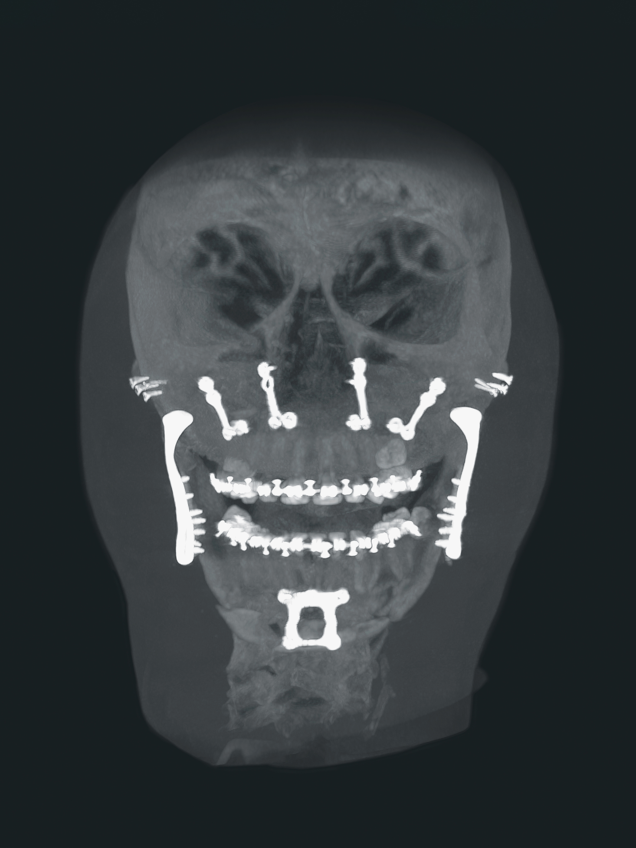
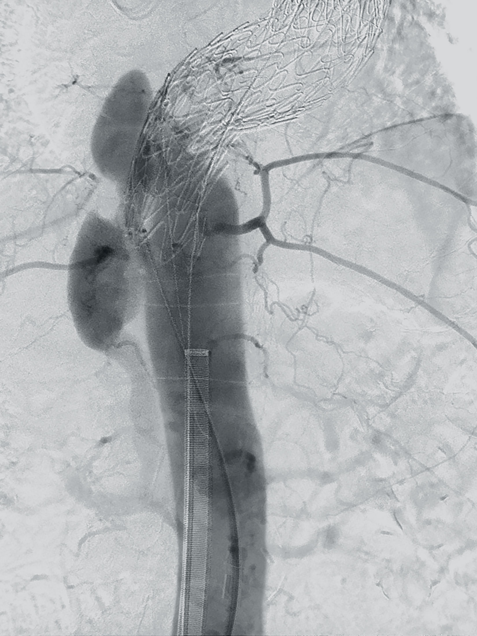
Pelvis surgery
“The Orthopedic Trauma Division at the University of Texas Health Science Center provides treatment for the full spectrum of orthopedic trauma. 3D imaging has been a ‘game changer’ for me and my colleagues. It has improved not only our technical capacities and judgment, but also our teaching of medical students and residents and our care of patients here in Houston. Misplaced screws can be corrected, suboptimal reductions can be adjusted – all without the dreaded delays in diagnosis, the need for revision surgeries, or other related problems. With intraoperative 3D imaging, treatments are more precise and patient safety increases.”
— Dr. Milton Routt
University of Texas Health Science Center, Houston, USA
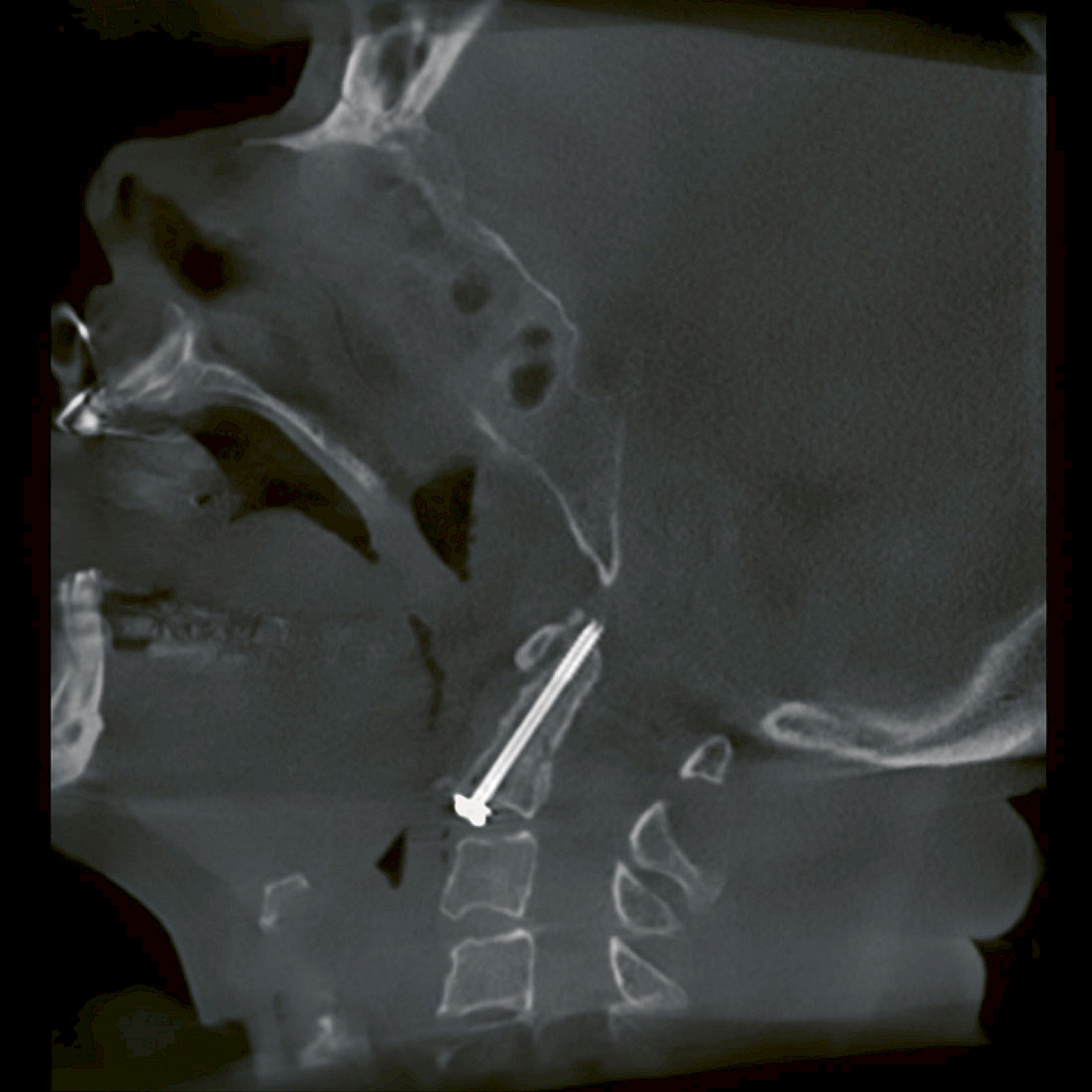
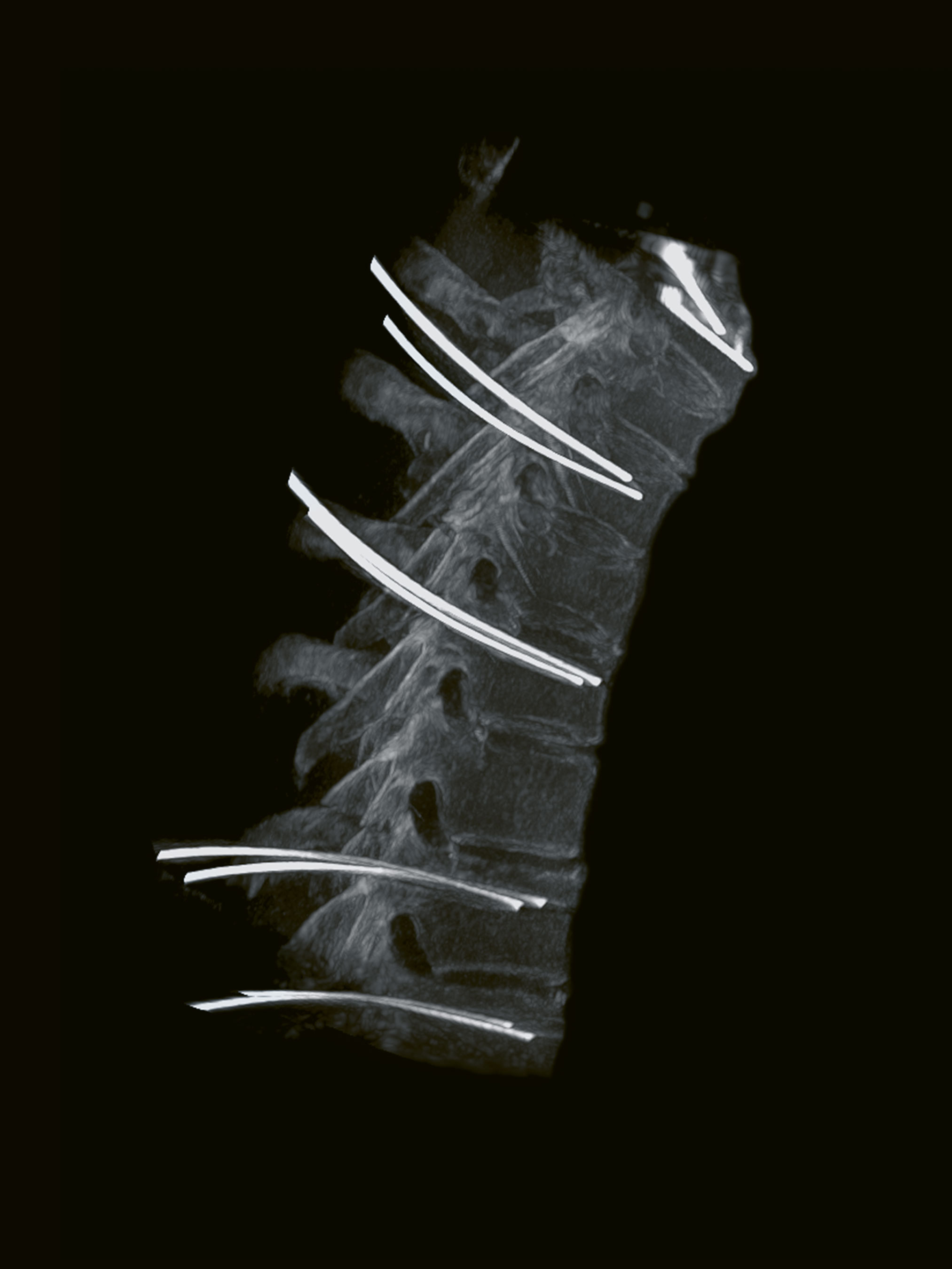
Spine surgery
“3D Imaging allows us to achieve high precision in complex surgical procedures. Moreover, with the ability to perform intraoperative control scans, we can considerably reduce the need for postoperative CT scans. With this 3D scan of a cervical spine there are significant dose savings compared to a CT examination. For me, dealing responsibly with radiation is one of the most important parameters when it comes to patient safety. For this reason, my team and I value the Low Dose Mode that helps us achieve exceptional clinical images with minimized radiation.”
— Prof. Christoph Josten
University Hospital Leipzig, Germany
Disclaimer
1
The CMOSline is a system configuration based on the Ziehm Imaging CMOS flat-panel detector.
More information on Ziehm Vision RFD 3D
Explore our image gallery on hand and foot surgery
Explore our image gallery on cochlear implantation and ENT surgery
Explore our image gallery on orthopedics and traumatology
Explore our image gallery on spinal surgery
—
This feature was published in issue 2.
Download issue 2 as PDF