Image of the Year
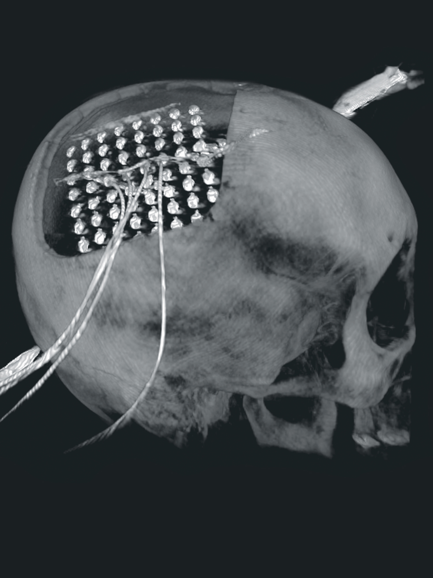
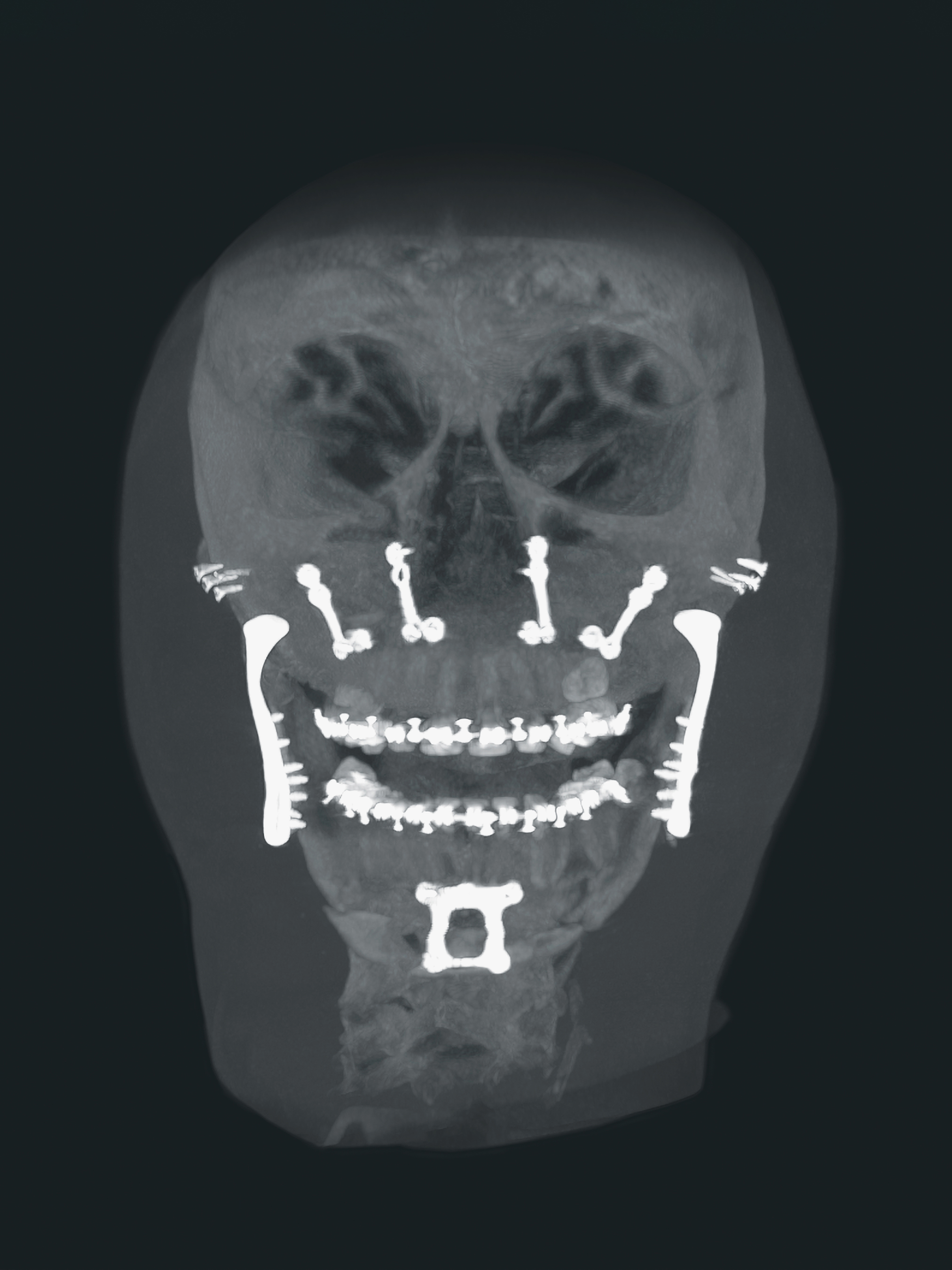
Acquired at the Medical University of Frankfurt, Frankfurt am Main, Germany, with a Ziehm Vision RFD 3D.
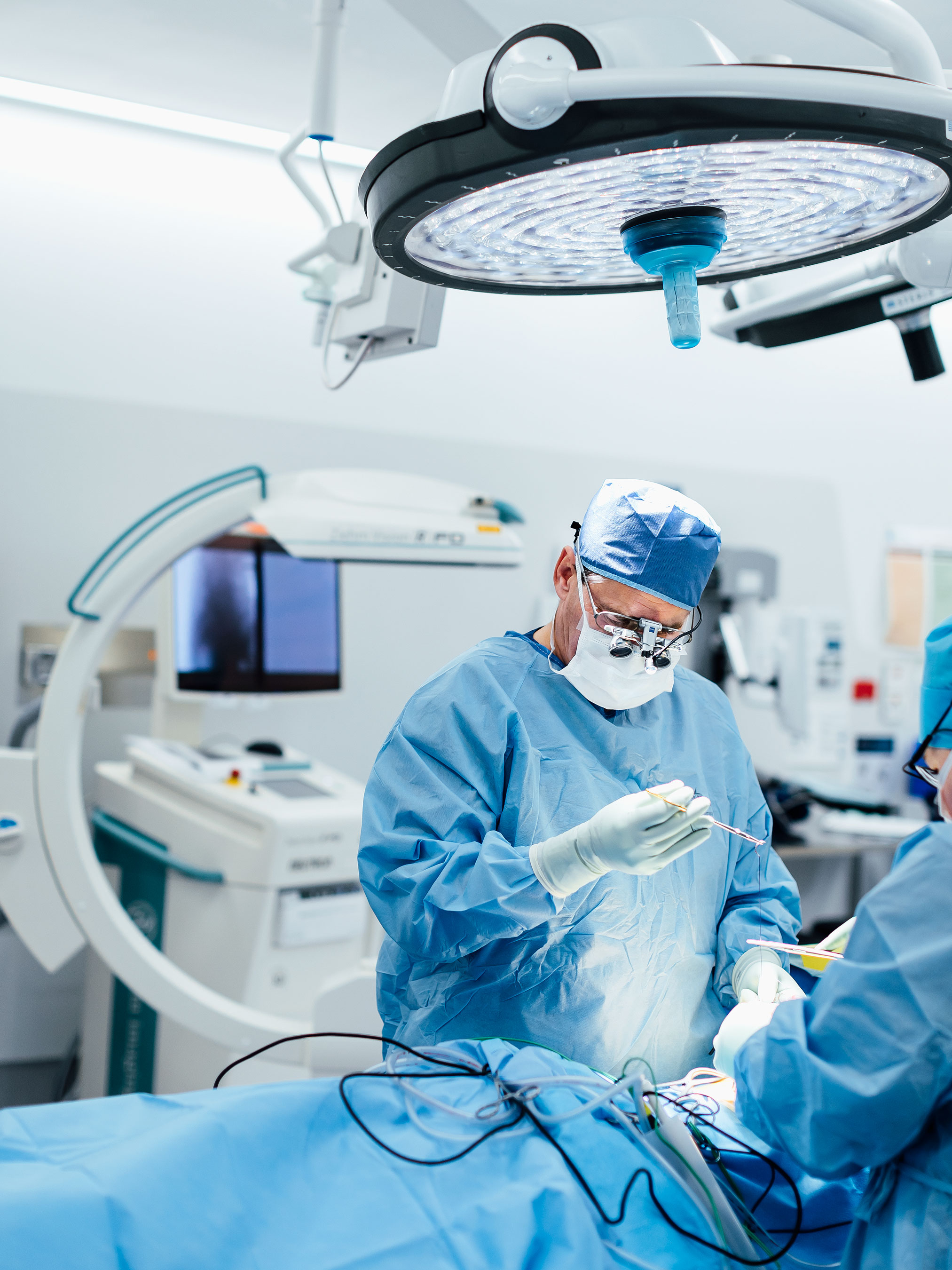
Nearly 50 million people worldwide suffer from epilepsy. For about a third of those affected, the only possible therapy is a neurosurgical procedure. In order to carry out a almost injury-free operation, stereotaxy is used. This means that several imaging techniques such as MRI, CT, robotic-guided navigation, and intraoperative X-ray all work together.
During the surgery, a grid of subdural electrodes is implanted that works similarly to a seismograph that measures earthquakes. The affected regions of the brain can be exactly plotted for subsequent therapy. Due to its CT-like image quality, the C-arm Ziehm Vision RFD 3D serves as an intraoperative control alternative, contributing to an optimum surgical outcome.