Synergies
Photos
Iona Dutz
The ongoing development of medical devices depends on cooperation between healthcare professionals and industrial developers. Prof. Dr. Christoph Josten tells us more about the role and trends of mobile imaging in trauma surgery at Leipzig University Hospital and about collaboration with Ziehm Imaging in these areas.

Professor Josten, what do you think sets the University Hospital in Leipzig apart?
Leipzig University Hospital is the second oldest university hospital in the German-speaking world. It has a very good reputation, which is also reflected in recurring awards. Eight years ago, I was faced with the challenge of merging the specialities of orthopedics and trauma surgery. I succeeded in building the Clinic for Orthopedics, Trauma Surgery and Plastic Surgery into one of the major university institutions in Germany, both in terms of patient numbers and the medical spectrum. The university hospital is certified as a Level 1 trauma center and has the highest level of certification as an endoprosthetics center as well as in spinal surgery. In addition, the clinic has very modern structures. The position of clinic director is not for life, as is common at most other universities in Germany. The executive director is elected by the five equally appointed professors of the Trauma Surgery, Orthopedics, Spine Surgery, Arthroscopy Sports Medicine, and Plastic Surgery departments on a regular basis, which strengthens the team spirit. I am proud that we have done so well in Leipzig so far.
Before you became medical director of Leipzig University Hospital, you worked in trauma surgery for 40 years. What is so appealing to you about this specialty?
Patients are often in life-threatening situations. You have to be able to make decisions quickly, and have good, broad-based medical knowledge and manual dexterity. I have never regretted my decision to become a trauma surgeon because of this fascinating combination.
What role does imaging play in trauma surgery?
Optimum imaging is indispensable in trauma surgery and an essential component of quality. Good, sharp images are important for making a diagnosis. But good imaging is also essential during the procedure to objectively assess surgical steps. After, or better, during the operation, good images can be used to check if the surgeon is satisfied with the result or whether he needs to make further adjustments.
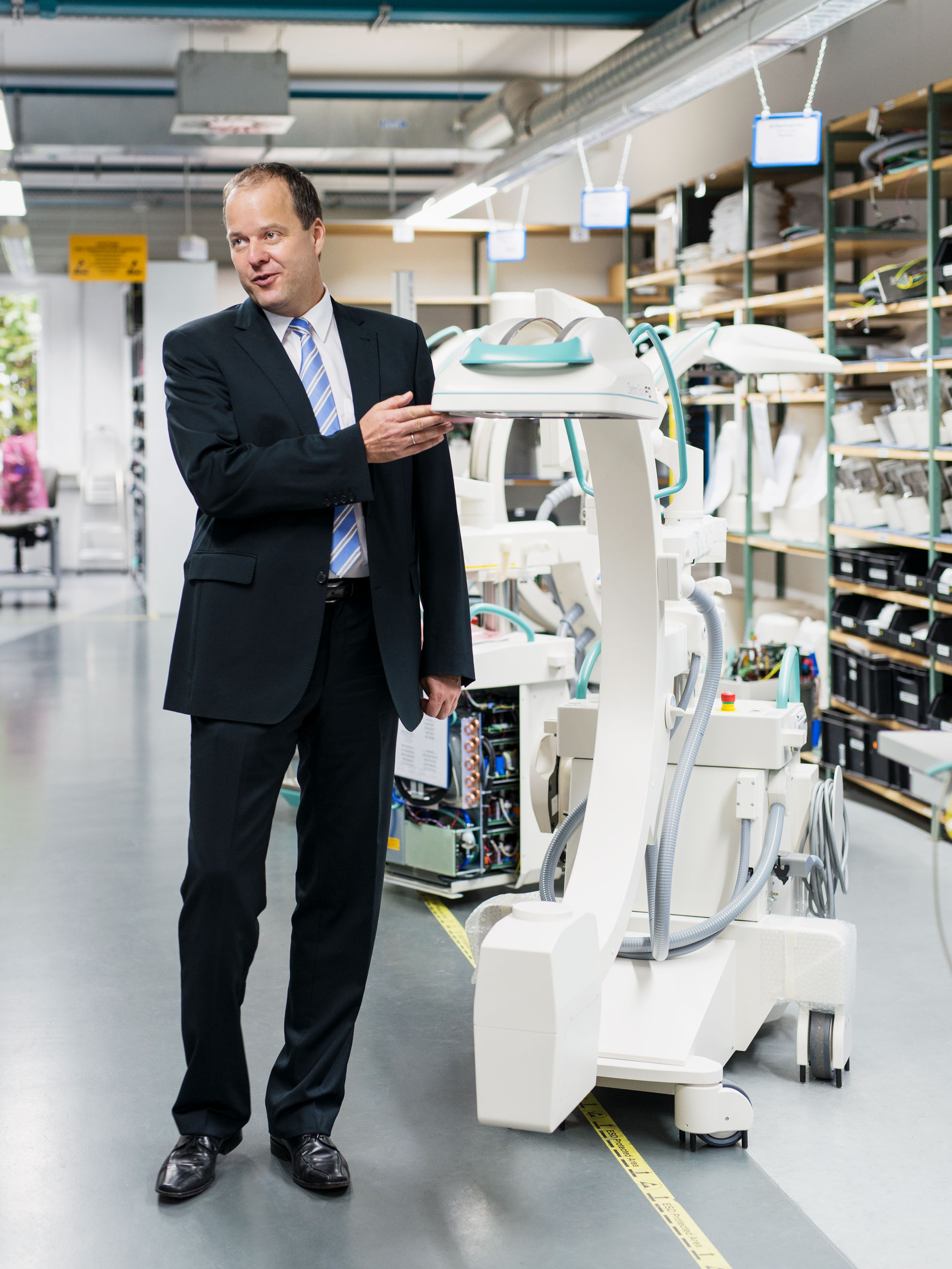
Why did you decide to use a mobile C-arm for imaging?
I’ll try to explain it like this: If I can get a car with four-wheel drive, I’ll take it. Since the 1980s, when the first mobile C-arm from Ziehm Imaging came on the market, it has been part of my daily surgical routine. Before that, when there was a fracture, usually an X-ray assistant had to come from the X-ray department. Many of those involved in the operation left the room, while some, protected by lead vests remained with the patient. X-rays were taken and stored on a cas vests sette. Worst case, this then had to be taken to the radiology department. It could take up to 15 minutes to get an image, which was sometimes underexposed or blurry. Intraoperative imaging has greatly improved this process. The mobile C-arm is brought into the OR, the images can be taken right away and displayed on the monitor.
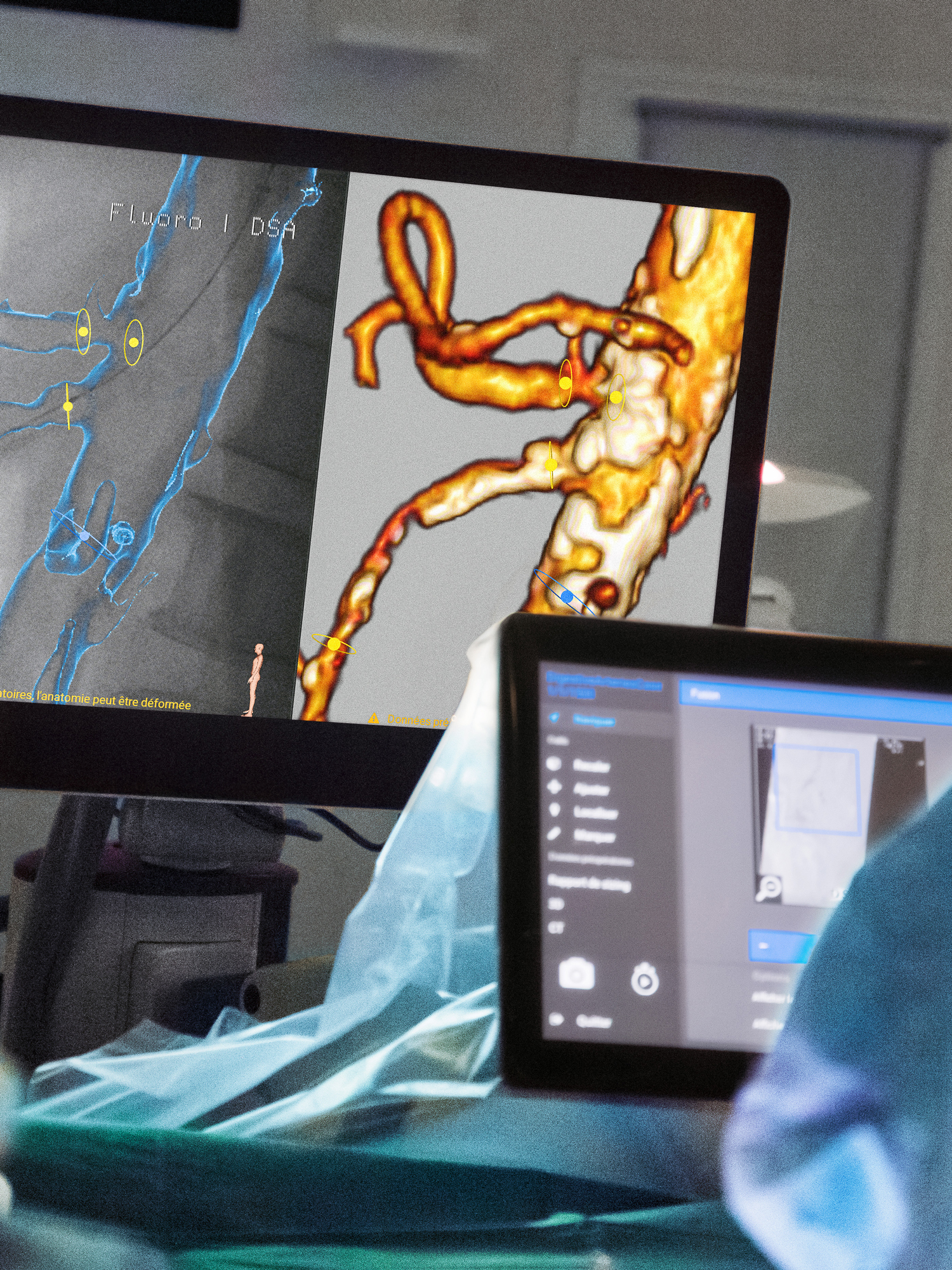
For a long time, 2D images were common in intraoperative imaging. Especially in orthopedics and trauma surgery, 3D imaging has been gaining in importance for years. What advantages does a three-dimensional image offer?
We were one of the first pilot clinics to work with 3D technology in imaging when it entered the market. It was like watching a movie with 3D glasses today. Thanks to the three-dimensionality, you dive into the anatomical structure in much more detail. With two-dimensional X-rays, you only see two planes. In between, however, there are umpteen degrees of angles that obscure something or that cannot be visualized properly. To get a usable image, you would often have to take countless X-rays. With the 3D scan, there is much less X-ray exposure and anatomical structures are much easier to see. This has helped me enormously, especially with complex fractures, to better understand the fracture and repositioning mechanism on the one hand and, of course, on the other, also to check the result after the operation. The 3D image is much more informative and meaningful because you can see all the levels of the joint and additional levels that are easier to miss in a 2D image. In my opinion, the advancement in 3D imaging has brought a huge increase in knowledge and a significant improvement in the quality of care.
You mentioned intraoperative control with the help of a 3D scan. How has this helped you with your work as a trauma surgeon?
I was one of the first trauma surgeons to argue that intraoperative 3D control should become a must. It gives you the opportunity to assess the outcome before the end of an operation and thus prevent follow-up surgical procedures, including anesthesia. After all, how do you explain to a patient at the X-ray check the next day that the surgical result is not optimum? That he must either accept the result with all its consequences or agree to another operation? Another argument in favor of intraoperative 3D scanning is the very good image quality. In the past, especially with complex fractures, repositioning maneuvers, and osteosyntheses, a postoperative CT scan was often necessary to check the result of the procedure. Today, 3D imaging is often so good that there is no longer any discernible difference in quality. In addition, the significantly lower radiation exposure benefits both the patient and the user. In my opinion, intraoperative 3D imaging is now an indispensable part of any advanced trauma surgery or orthopedics OR.
How do you think 3D imaging will change in the future?
If the focus of imaging systems is on the quality of imaging, reduction of radiation exposure, as well as ease of handling, and this is expanded to include digitally networked systems, there is enormous potential for development. In the future, it may also be possible to better image soft tissues and vessels. I still see potential in these areas to enable the progress of medical specialties beyond trauma surgery.
It is also possible to perform navigated procedures with C-arms from Ziehm Imaging. How important was navigation to you during operations?
I was a fan of navigation from very early on. I started doing CT-based navigation about 20 years ago. At that time, it was still very cumbersome, raised a lot of questions, was timeconsuming and not of sufficient quality. I decided at that time to wait for further progress in the technology. When image intensifier-based navigation came on the market in the mid-2000s, I started again, and I quickly realized that the development was a quantum leap. The advantages of 3D imaging that we just discussed could then be combined with navigation. The accuracy was excellent. From then on, it was possible to perform far more minimally invasive surgeries and reduce surgical dimensions. On top of that, the image size expanded thanks to the introduction of the flat panel. With the larger images, it was possible to navigate well and safely in more complex, larger body regions. Since then, it has been a must for me to perform certain procedures with navigation. One needs significantly fewer postoperative control CTs, since the 3D images obtained intraoperatively with the mobile C-arm from Ziehm Imaging offer good image quality. Nevertheless, in my opinion, it is important to be able to master the surgical challenges without navigation and to be technically versed in the surgical repertoire so that it’s possible to operate without navigation at any time if necessary.
How do you see the market for navigation and robotics?
We, as a hospital, are sure that the advances in imaging, navigation, and computer-assisted surgery are unstoppable, as they lead to a huge improvement in the quality of many invasive procedures. That’s why we established the ‘Center for Computer Assisted and Navigated Surgery’ last year. We are also getting three state-of-the-art operating rooms this year, which will be equipped with the latest CT and 3D-assisted navigation systems. The quality standards of surgeries are so high that without such systems it is no longer possible to meet the demands of today’s high-tech medicine.
Do you think navigation will continue to gain in importance in the future?
You can compare developments in this area with road traffic. In the past, many people claimed that they didn’t need a navigation system in their car because they had a strong sense of direction. Today, almost no one drives without one. People know that they might not need it every day, but if the route is unknown or they are looking for the fastest route, they inevitably fall back on navigation. It’s the same with navigation in the operating room. That is why I believe that there is still potential in many areas where it is not yet considered today. Since navigation systems deliver an improvement in quality, they will become even more important.
Let’s take another look at the past. When and why did you decide to work with Ziehm Imaging fluoroscopy systems?
It was so long ago that I can’t really say – certainly more than 30 years ago. I noticed Ziehm Imaging mainly because the equipment was much handier than others on the market. Since space in the operating room is usually very limited, this criterion was and remains important. In addition, Ziehm Imaging offers user-friendly fluoroscopy systems. But of course, the very good image quality is the main thing. I’m also convinced by the good operability, the easy maneuverability, and space-saving.
You have worked a lot with Ziehm Imaging in recent years. What can you tell us about your collaboration?
When an innovative company like Ziehm Imaging meets physicians who are interested in new developments, a constructive collaboration almost inevitably results. In my experience, Ziehm Imaging is very open to feedback from physicians about their systems and the resulting needs and wishes. At an early stage, we decided on development projects with Ziehm Imaging and conducted clinical and anatomical studies to find out, for example, how to improve image quality and reduce radiation exposure at the same time. A trusting and result-oriented collaboration like this gives rise to new projects, such as systems that will ultimately be launched on the market for the benefit of the patients.
What added value do you see in working with Ziehm Imaging?
For me, the most important aspects in my direct collaboration with Ziehm Imaging were the technical progress as well as the practice-oriented applicability and, ultimately, the benefit for the patient. On the other hand, the international contacts that were established through the collaboration and that then developed into global partnerships were also important. Guest physicians came from a wide variety of countries and were able to see the technical possibilities of the systems for themselves. We were able to train doctors here in Leipzig, which also led to an extensive medical exchange. This was very important, especially for me as a scientific university lecturer. These are all very positive aspects of the collaboration between medicine and industry. Not only are products developed that benefit the patient, but these products are also accepted internationally and scientifically. This contributes to the reputation of Germany as a center of manufacturing and science.
You are now the Medical Director of Leipzig University Hospital. How has your collaboration with Ziehm Imaging changed in this role?
Due to my current position, I no longer work personally with Ziehm Imaging. However, Leipzig University Hospital is still a partner. Of course, I monitor developments, keep in touch with medical colleagues and users, and make sure that they have the best possible equipment for the best possible therapy for our patients. And I hope that Ziehm Imaging will continue to dedicate itself to research and new developments in exchange with medical professionals, even without my specific involvement.
What opportunities do you see in the future for Ziehm Imaging’s collaboration with Leipzig University Hospital?
On the one hand, the business relationship continues, as we have Ziehm Imaging equipment in our hospital. That means the cooperation will continue. But of course, I would also like to see scientific collaborations in the future with the colleagues who now run the Clinic for Orthopedics, Trauma Surgery and Plastic Surgery. I would also like to see companies getting involved in our new center for robot-assisted and navigated surgery. After all, establishing a leading international center for technically-assisted surgery can only succeed through good cooperation. Both sides must approach each other, i.e., the industry, the physicians, and the clinics must approach the manufacturers so that joint projects can be realized. That’s why I’m sure that there will be fruitful cooperation in the future as well.
And finally, a look into the future: How do you envision the optimum clinical workflow in ten years?
My vision of a future surgical workflow could perhaps best be compared to autonomous driving. You sit in a car that drives itself based on navigation. It warns you when there is a risk of traffic jams, brakes automatically, and never drives too fast, so that the driver only has to intervene if he doesn’t trust the automatic system or gets into an exceptional situation. Similarly, I can also imagine future interventions consisting of a combination of robotics and navigation. Certain surgical steps would take place fully automatically and surgeons would only have to intervene at critical points, and perhaps not even necessarily be on site in the operating room.
More information on Leipzig University Hospital
This interview has been pulished in issue 5 (2022).
Download issue 5 as PDF