Radiation protection
in pediatrics
Children have a higher risk of developing cancer after exposure to radiation than adults. This makes it even more important for physicians to minimize radiation exposure for children, insofar as it is compatible with meaningful diagnostics and therapy. We, as manufacturers of X-ray systems, can aid hospital staff in their everyday work with hardware and software solutions to contribute to this end.
1
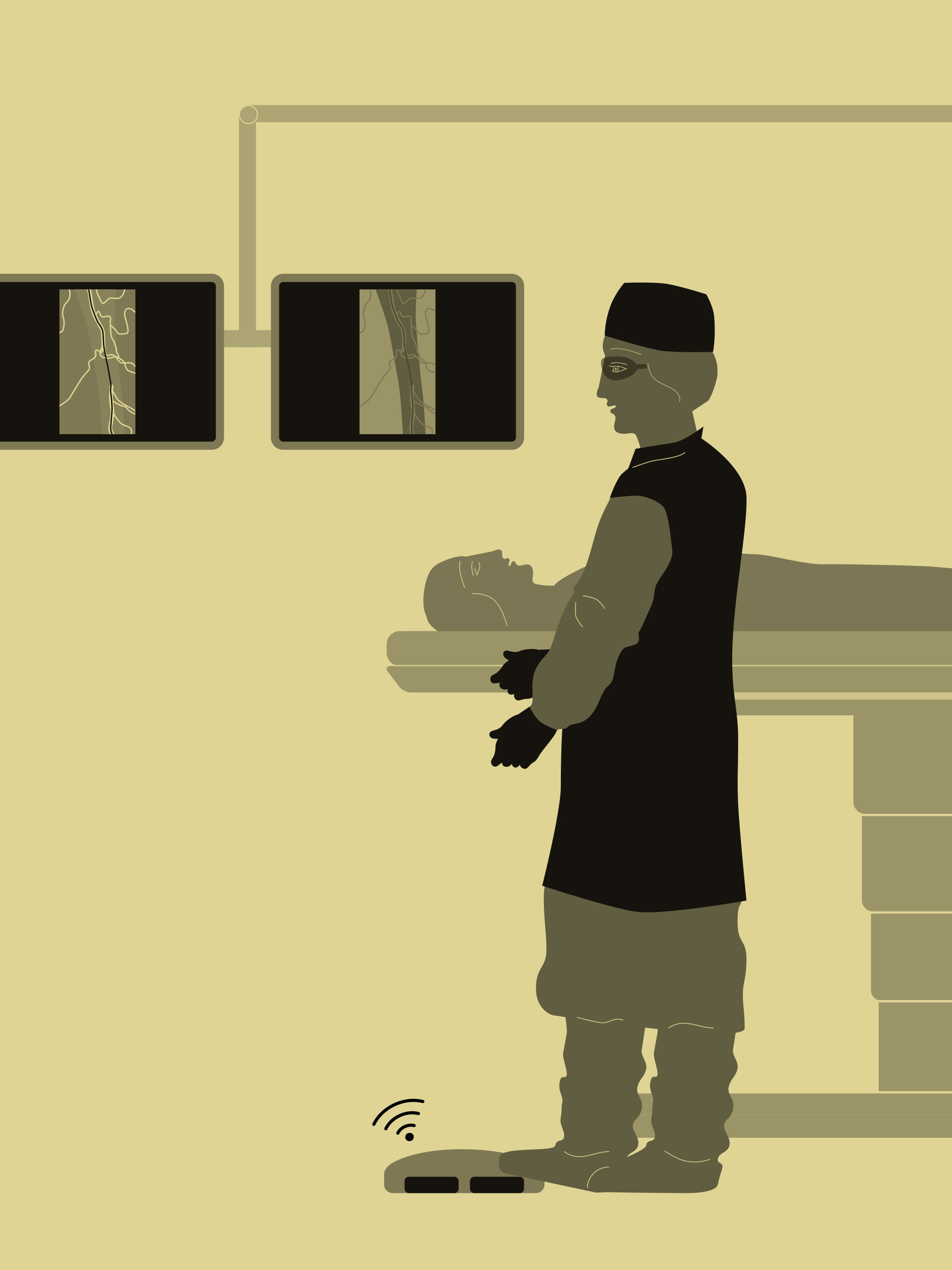
Mastering the basics of X-rays
Apply the ALARA principle
‘As low as reasonably achievable,’ or ALARA, is a global radiation protection principle for the optimized use of radiation. Basically, it means that radiation should be used as judiciously as possible on humans, animals, and materials. ‘Judiciously’ means that every dose should be as low as possible, considering all advantages and disadvantages. Especially in pediatrics, the ALARA principle is of highest priority.
Avoiding unnecessary radiation
With every X-ray, but especially when imaging children, it is important to use radiation with special care. This means that radiation should be conducive for the success of the therapy goals: Is fluoroscopy absolutely necessary? Are there alternatives, such as the possibility of radiation-free magnetic resonance imaging or ultrasound? The answers to these questions must be weighed carefully.
Protect all sensitive areas
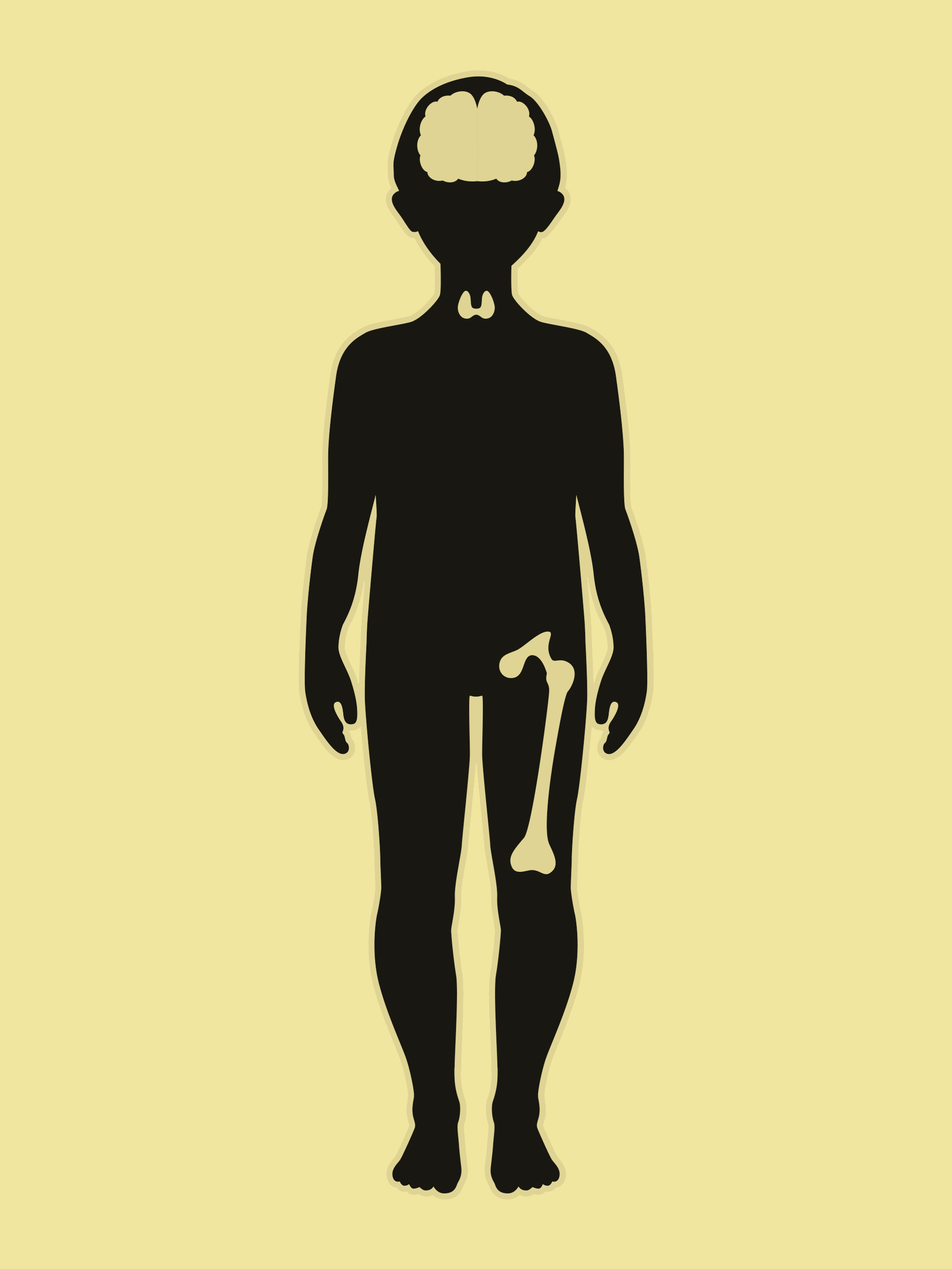
The region of interest (ROI) is the area of relevance to be examined. It should always be as small as possible, because this keeps radiation exposure as low as possible. Everything outside the ROI is shielded by special lead blankets. In children, skin, brain, eyes, thyroid and gonad glands, as well as breast and bone marrow should be especially protected.
Keep fluoroscopy exposure times brief
If radiation has to be used, exposure should be as brief as possible. With current technology, some presettings of the system can be changed without X-ray exposure. This allows targeted radiation, and the fluoroscopy exposure time is limited.
Working close to the detector
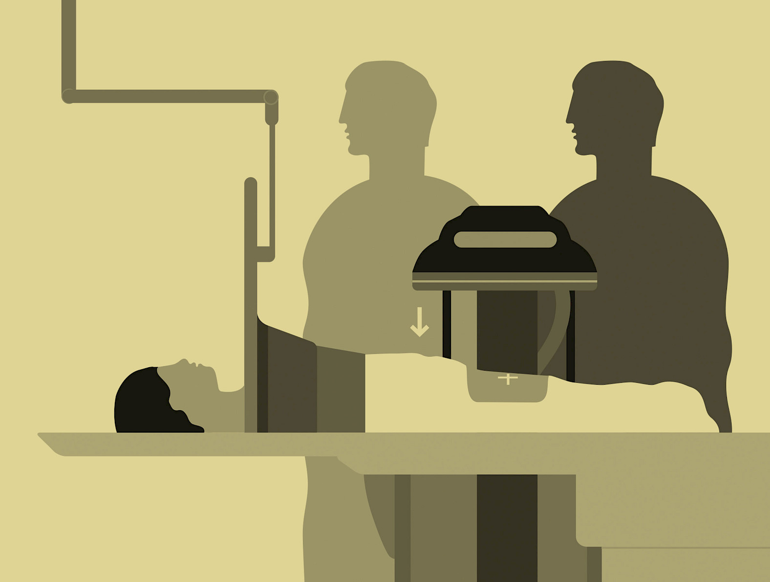
Positioning the detector as close to the patient as possible has several advantages: image quality is better, the dose is reduced, and the field of view (FOV) is extended. Scattered radiation is also significantly reduced, which is beneficial for the staff, in particular.
2
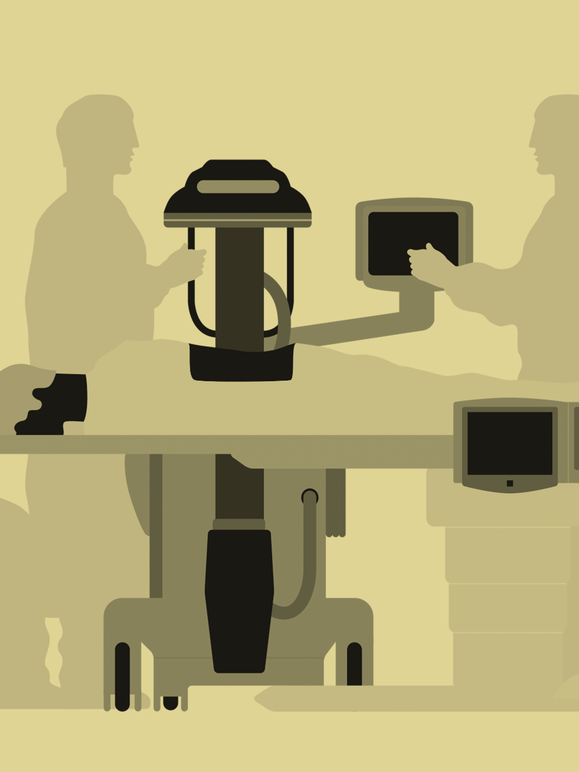
Correct use of the latest technology
Pulsed fluoroscopy
In some countries, pulsed fluoroscopy is standard for pediatric fluoroscopy due to legal requirements. However, the correct application with individual reduction of the pulse rates is important. Thanks to new Ziehm technologies, image quality comparable to that achieved with high pulse rates can be acquired at low pulse rates.
Using innovative technologies
Pediatric procedures should be carried out with the latest technology – in recent years, aspects such as dose management, and applications supporting ease of use and image-quality optimization have been continuously improved. Ziehm C-arms not only deliver very good image quality, but also improved copper and carbon pre-filtering. This is responsible for increased hardening of the X-ray beam, which in turn leads to a reduced skin entrance dose. In addition, all Ziehm Imaging CMOSline devices come with a ‘Low Dose Mode’ presetting.

3
Optimizing C-arm settings
Radiation-free patient positioning
The laser can help determine the region of interest (ROI) without using any radiation. The C-arm is thus positioned quickly and optimally without additional dose. The virtual collimators can be set asymmetrically to the ROI on the touch screen with just one hand. The smallest possible radiation field is then displayed for capturing the image.
Using special anatomical programs
Ziehm C-arms offer various so-called ‘anatomical programs’ (APR) for each important body region. These programs are optimized for the respective area in terms of image quality and dose. In addition to the APRs, patient-specific options can be selected with modifiers. The modifiers can also be used to activate further dose-reducing options for children. In addition, there is a low-dose button that is always activated during a pediatric examination.
Applying magnification modes
In the so-called ‘MagModes,’ an enlarged view of the relevant anatomy can be achieved at the same dose. Another advantage of the magnification modes is the irradiation of a small area. The higher resolution and the lower noise of the CMOS detector also allow a more precise image without increasing the dose.
Dilute contrast medium
If a contrast medium has to be used, high image quality can also be achieved with diluted contrast medium. This reduces the strain on the body.
Removing the anti-scatter grid
Due to their smaller anatomies, the scattered radiation created during fluoroscopy of children or other small patients is significantly lower. Therefore, the anti-scatter grid can be removed for pediatric or other dose-sensitive applications. Nevertheless, excellent image quality can be achieved with a significantly lower dose.
These tips & tricks were published in issue 4 (2020).
Download issue 4 as PDF