Three steps
to the perfect
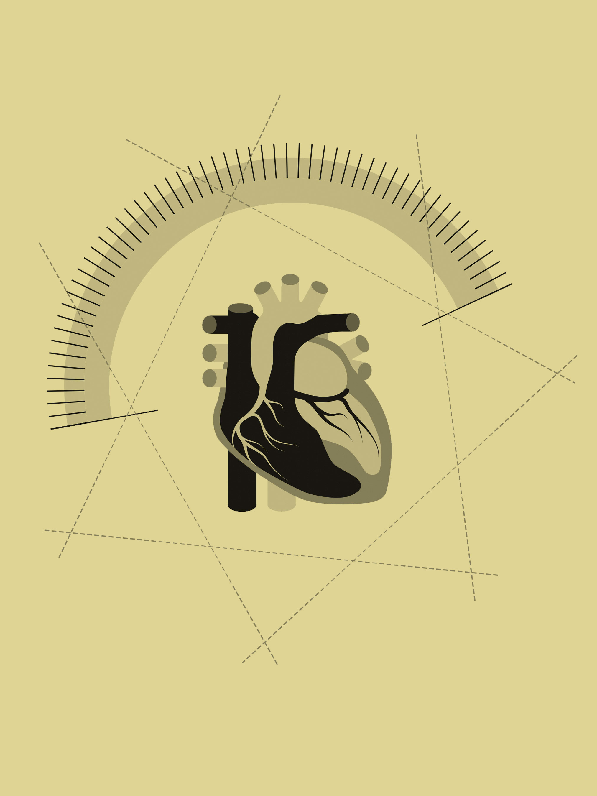
vascular image
Imaging blood vessels, aneurysms and stents is a daily procedure at many hospitals worldwide, in both standard and highly complex hybrid operating rooms. At Ziehm Imaging, Product Managers Jörg Leonhardt, Wolfgang Keller, and Florian Schnabel work hard every day to create the ideal conditions for demanding vascular operations with a mobile C‑arm. They share their knowledge about image quality and reducing radiation dose as well as optimized surgical procedures with physicians and hospital staff worldwide.

1
Setting up
the best surgical environment
Proper radiation exposure protection
In addition to a ceiling-mounted exposure protection panel made of lead acrylic, the ideal protection for personnel requires radiation protection attached below the table. The OR staff should wear not only the classic exposure protection vests and gowns, but also protective glasses or visors in addition to special gloves and hoods.
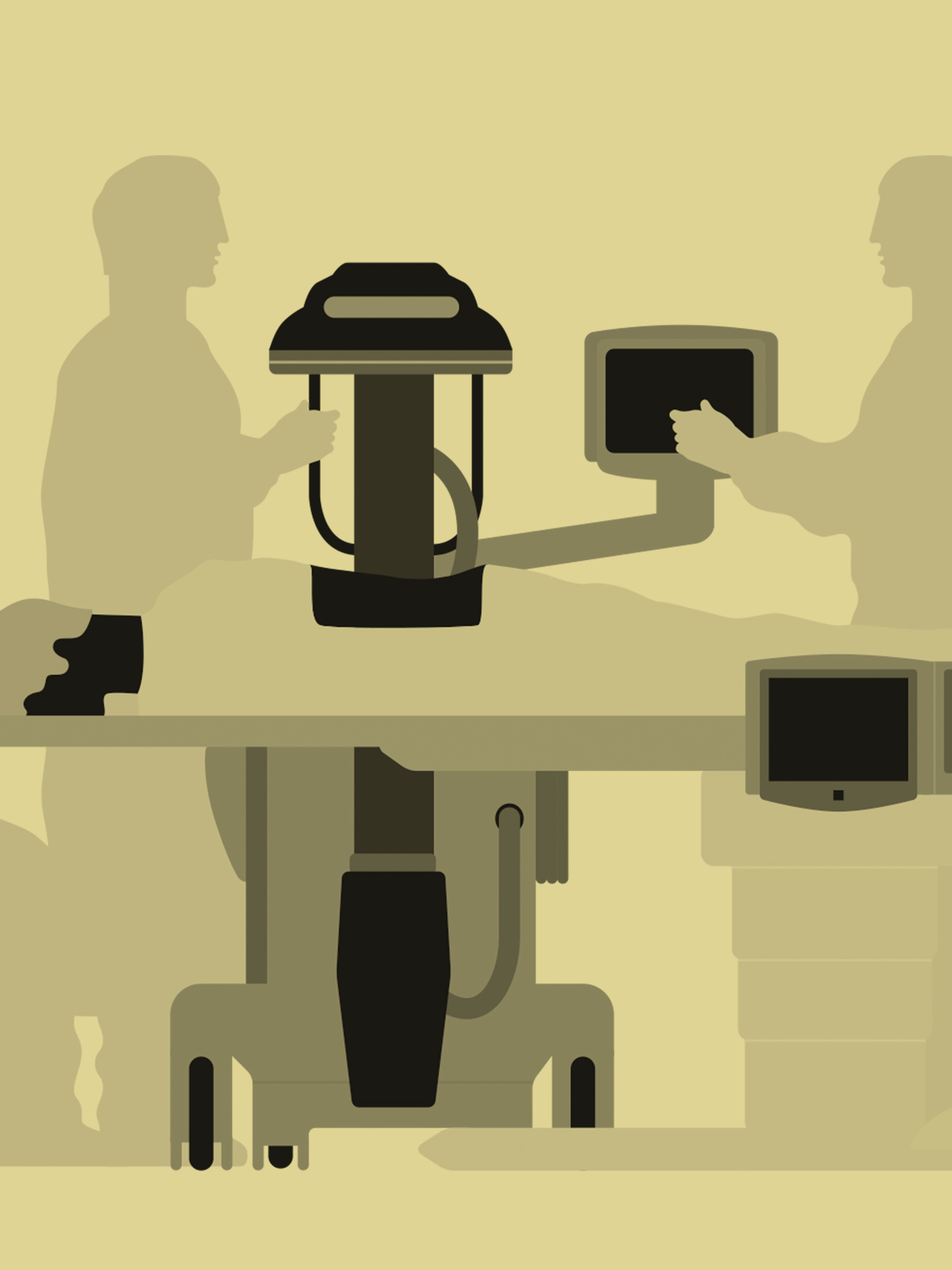
The proper operating table
A floating, X-ray translucent, carbon table element is ideal. With just one hand, the patient can be brought into the best position with respect to the C‑arm in all four directions. The table top can also be imaged without creating any artifacts.
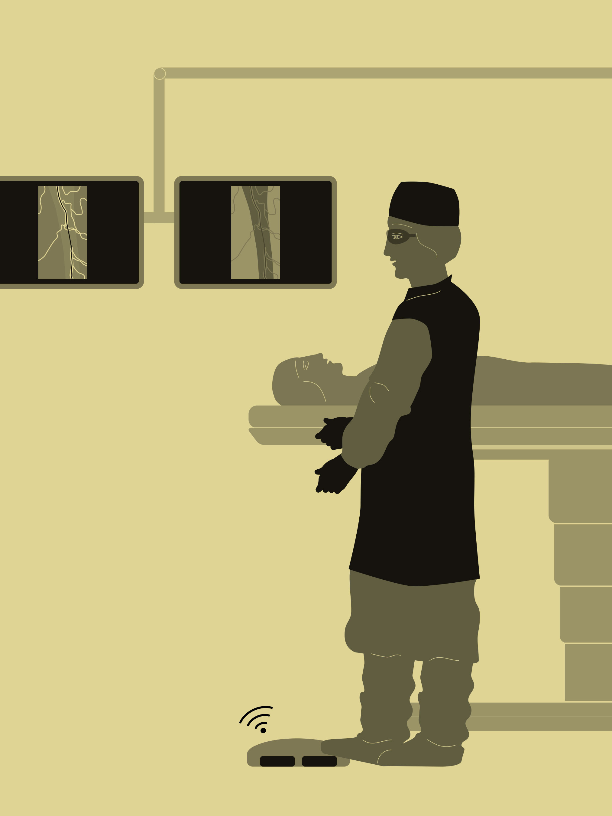
Optimized system usability
There are two control modules that enable the physician to steer all functions of the C-arm. One is a touchscreen that operates the whole system. The other is a joystick that controls the motorization. Both control modules can be mounted in the sterile field — either on the table or on a special trolley. The wireless footswitch can be placed within easy reach of the operator, enabling intuitive and customizable use.
2
Important information
for vascular procedures
Positioning the system properly
The C‑arm should be positioned opposite the surgeon to ensure optimum freedom of movement. To reduce scatter radiation and maximize the field of view (FOV), the patient should be positioned as close to the detector as possible.
Accurate system positioning
The laser can be used to set the region of interest (ROI) without exposure, so the C‑arm can be positioned ideally and quickly, without additional dose. The table top floats, so the system doesn’t have to be moved. With the assistance of collimators along the course of the blood vessel, the smallest possible radiation field is displayed.
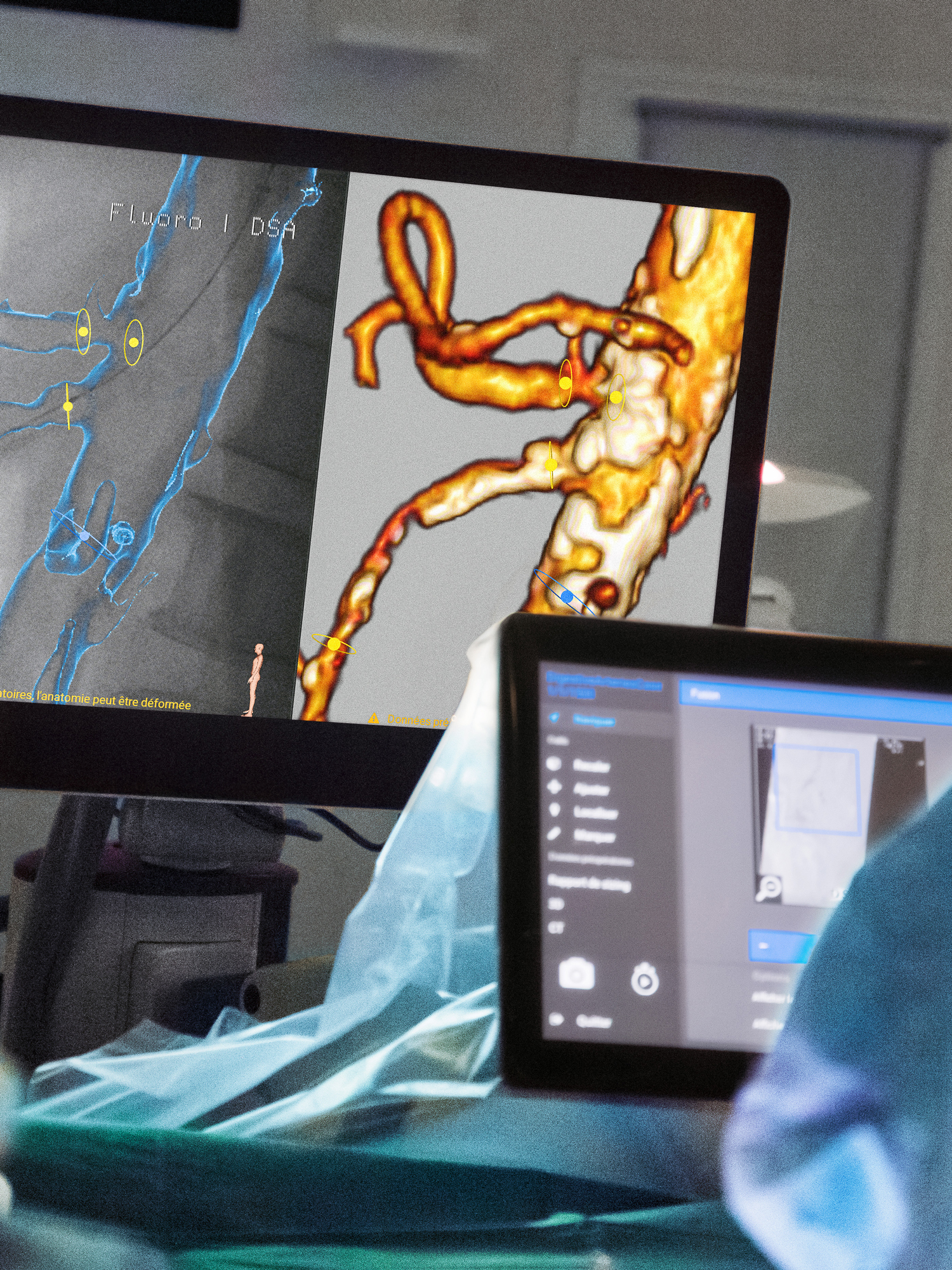
Using the Anatomical Marking Tool (AMT)
The AMT makes it easy to save markings and notes onto the live images: e.g. left/right side marking, drawings of blood vessels, anatomical landmarks or implant positions. This also makes the use of a contrast agent unnecessary.
Intuitive workflow with SmartVascular
SmartVascular enables independent switching among fluoroscopy, digital subtraction angiography (DSA), and roadmapping. The physicians can operate the system with the wireless footswitch with its customizable configuration. Further functions can also be accessed via touchscreen directly from the sterile field with one click.
3
The best tips
for complex vascular procedures
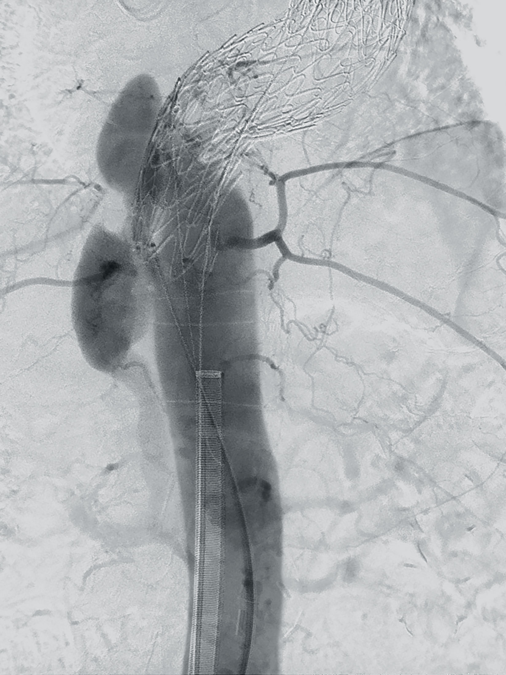
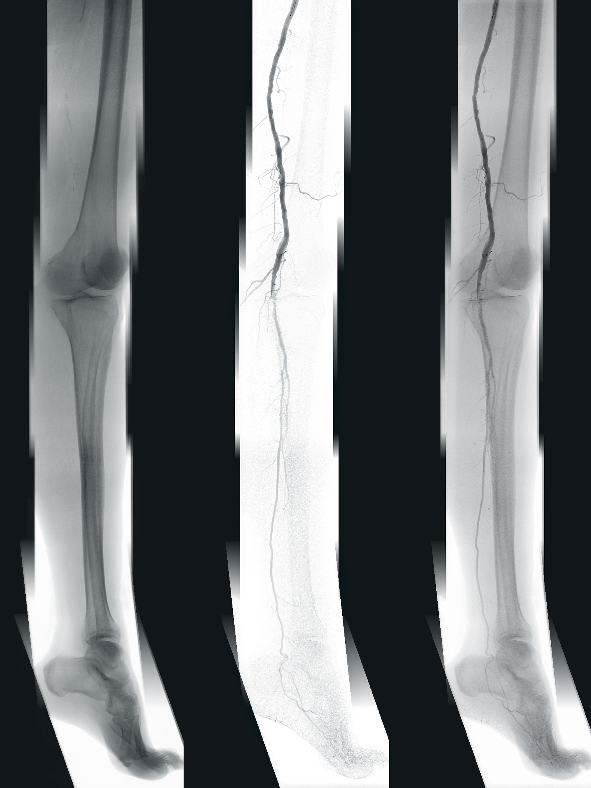
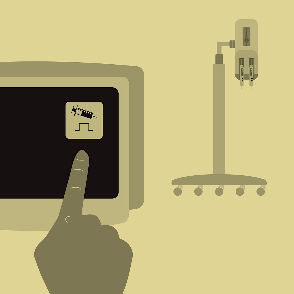
Using contrast agent
To make vessels visible during X‑ray monitoring, a contrast agent is injected. To improve effectiveness and patient comfort, the contrast agent can be diluted. To obtain the best image quality, even in complicated procedures, the C‑arm automatically triggers the injector for the contrast agent.
Selecting the right contrast agent
For patients for whom a conventional contrast agent cannot be used due to contraindications, CO₂ represents an advanced, safe, and cost-efficient alternative. CO₂ can also be used in a diluted state to keep the stress on the body as low as possible.
Advantages of CO₂ as a contrast agent
With the special CO₂ package, the imaging adapts perfectly to changed conditions. However, all of the workflows and tools remain unchanged for the user, so it appears that an immediate inversion of the subtraction image has occurred. The surgeon sees a familiar image — the CO₂ in the blood vessels is shown in black; the contrast and image quality remain unchanged.
Single frame roadmapping
If an image is less than optimum due to patient movement during the DSA, single frame roadmapping can be applied. In this case, the entire DSA sequence is no longer used for roadmapping, but instead, only the best image. This effectively eliminates the movement artifacts in the sequences, so the DSA with the injection of contrast agent doesn’t need to be repeated.
Breathing stop for better image quality
To keep patient movement to a minimum during the DSA and get the best possible image quality without movement artifacts for the subtraction, breathing can be interrupted. If this is known in advance, the patient can be hyperoxygenated beforehand.
These tips & tricks were published in issue 3 (2019).
Download issue 3 as PDF