Panorama
Clinical images
Giessen and Marburg University Hospital, Germany
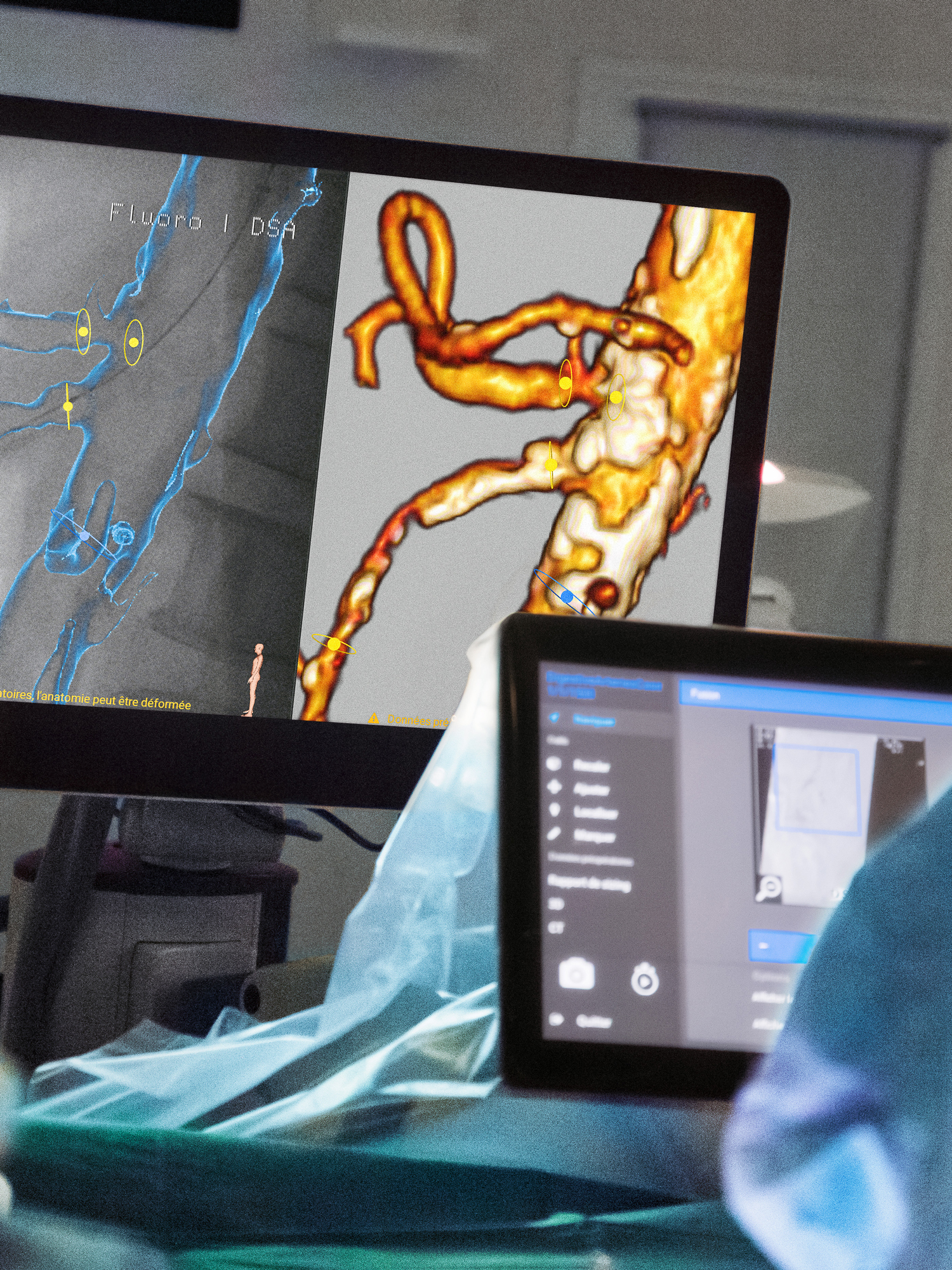
Complex endovascular procedures are performed in our mobile hybrid rooms all over the world. The team of vascular surgeons at Giessen University Hospital, Germany, uses the Ziehm Vision RFD Hybrid Edition mobile C-arm with the EndoNaut workstation. The intraoperative navigation system supports them during their procedures and provides them with a constant overview in 2D panoramic images of the extremities.
As the global population ages, the importance of endovascular therapies continues to grow. Studies show that approximately from 3 to 10% of the population in industrialized countries suffer from peripheral arterial disease (PAD). In 90% of cases, the disease affects the legs. In more than 95% of cases, the disease is caused by arteriosclerosis in which the narrowing of the arteries severely restricts blood flow to the extremities. Some may experience little or no discomfort while blood flow is still adequate in the early stages of the disease, but as it progresses, they may experience increasing pain in the calf while walking. Often, they must stop until the pain subsides before they can continue walking. PAD is also known as ‘window shopper’s disease’ (intermittent claudication) because people with PAD often stop to, supposedly, look in shop windows while walking through town. If PAD is not treated consistently, it can develop into gangrene requiring an amputation.
The probability of developing the disease increases with age. From the age of 70 onwards, the prevalence is around 15 to 20%. Classic risk factors, in addition to age, include nicotine abuse, arterial hypertension, hyperlipidemia, hypercholesterolemia, and diabetes mellitus.
Vascuar surgery
at Giessen University Hospital
The team of vascular surgeons at Giessen University Hospital is specialized in complex open surgical therapies and endovascular treatment of PAD, and uses both procedures for the maximum benefit of their patients. Prof. Dr. Johannes Kalder has been the head of vascular surgery since 2020. He and his team regularly operate on patients with PAD, performing an average of 350 endovascular procedures on the vessels of the lower extremities each year.
Today, Prof. Kalder and his colleagues PD Dr. Paula Keschenau, chief physician for vascular surgery, and Anne Lindemann, resident physician for vascular surgery, are operating on just such a case. The patient presented with stage IIb to III PAD. He could only walk 15 meters without symptoms, and was woken up at night by the pain in his foot. He had a stent implanted in his leg for PAD at another clinic years earlier.
The team has been using the Ziehm Vision RFD Hybrid Edition¹ since 2020, and the EndoNaut vascular navigation system² for endovascular treatment since 2022. The EndoNaut workstation is also important for this case. To localize the lesion, the team creates preoperative panoramic images with the EndoNaut workstation. The C-arm generates fluoroscopic and angiographic images that are transferred to the EndoNaut workstation.
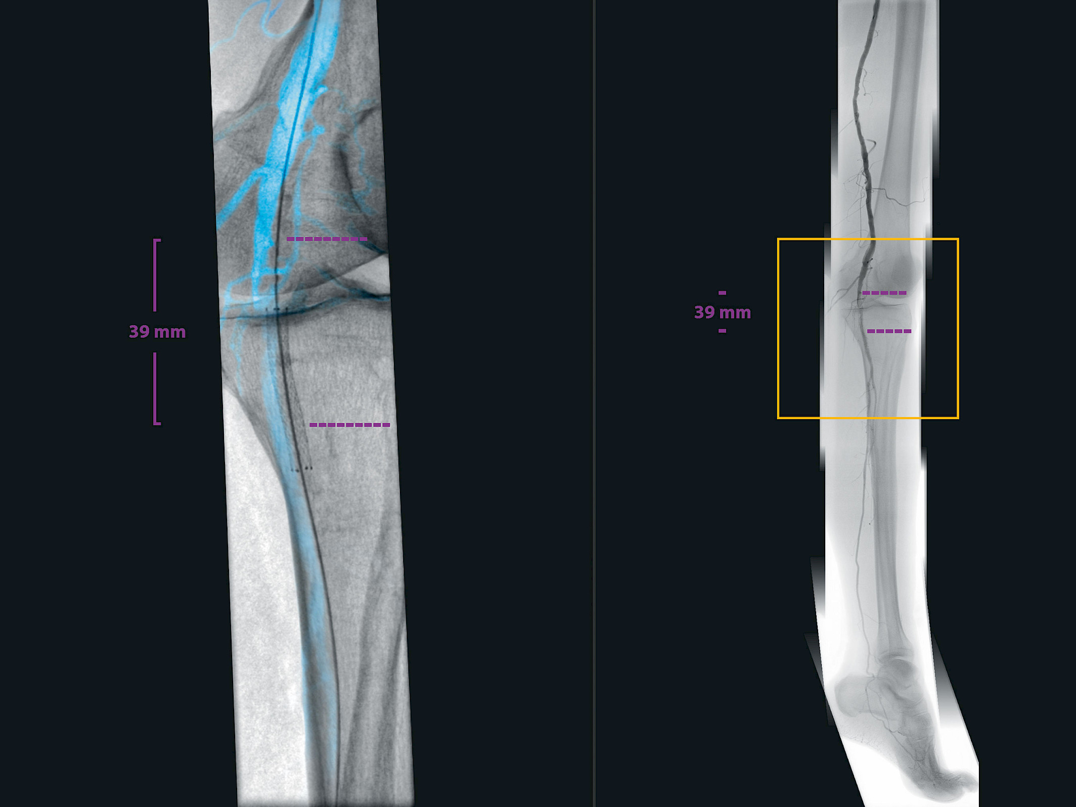
Determine the location of the lesion
“The combined panorama with the underlying bone makes it easier to determine the location of the lesion and optimally align the C-arm for the intervention.”
Corresponding images are stitched together to create the intraoperative 2D panorama. A fluoroscopic panorama, an angiographic panorama, and a combined fluoroscopic and angiographic panorama are automatically generated. “The combined panorama with bone overlay makes it easier for us to determine the position of the lesion,” confirms Prof. Kalder. “With this information, we can optimally align the C-arm for the intervention.” Dr. Keschenau adds that in the case of multiple lesions, it is also possible to avoid repeated angiography of individual segments and to target the lesions directly in the panorama. The system algorithm allows the creation of a very precise panorama, using automatic, near real-time 2D-2D registration between adjacent images with minimal overlap. The team is impressed by the good overview the panorama provides. “We need to perform fewer angiographies, and that means less contrast media use and X-ray dose.”
Target individual lesions
“If there are several lesions to treat, you don’t have to acquire a new angiographic image each time. Thanks to the panorama, we can target the individual lesions that have been marked and measured.”
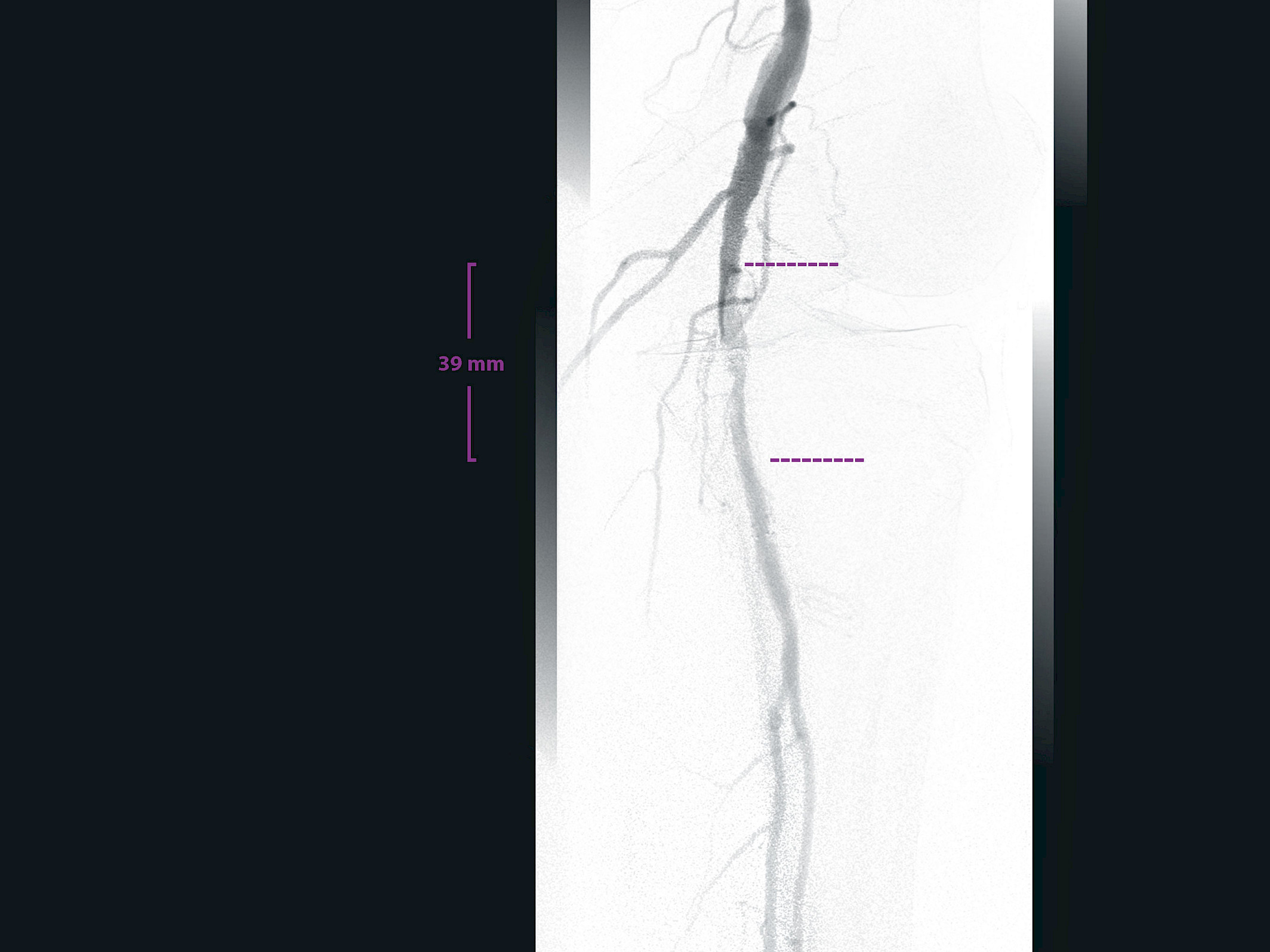
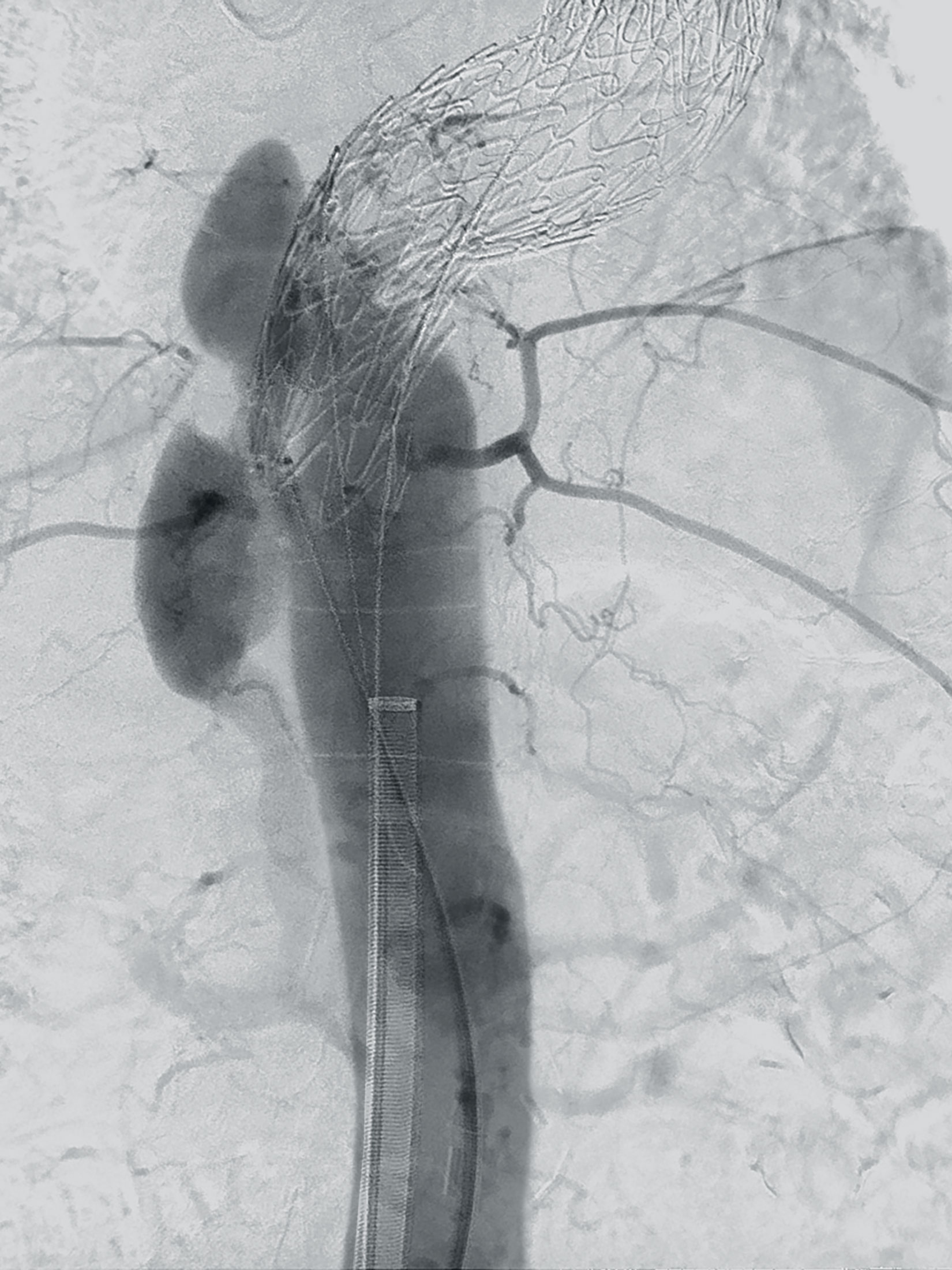
The lesions can be localized and marked on the panorama. This time the stenosis is in the popliteal artery. The EndoNaut workstation calibration function makes it possible to measure the length of the lesion to select the best balloon or stent. With great skill and the help of the navigation function and digital zoom of the EndoNaut workstation, the guidewire is successfully passed through the occlusion.
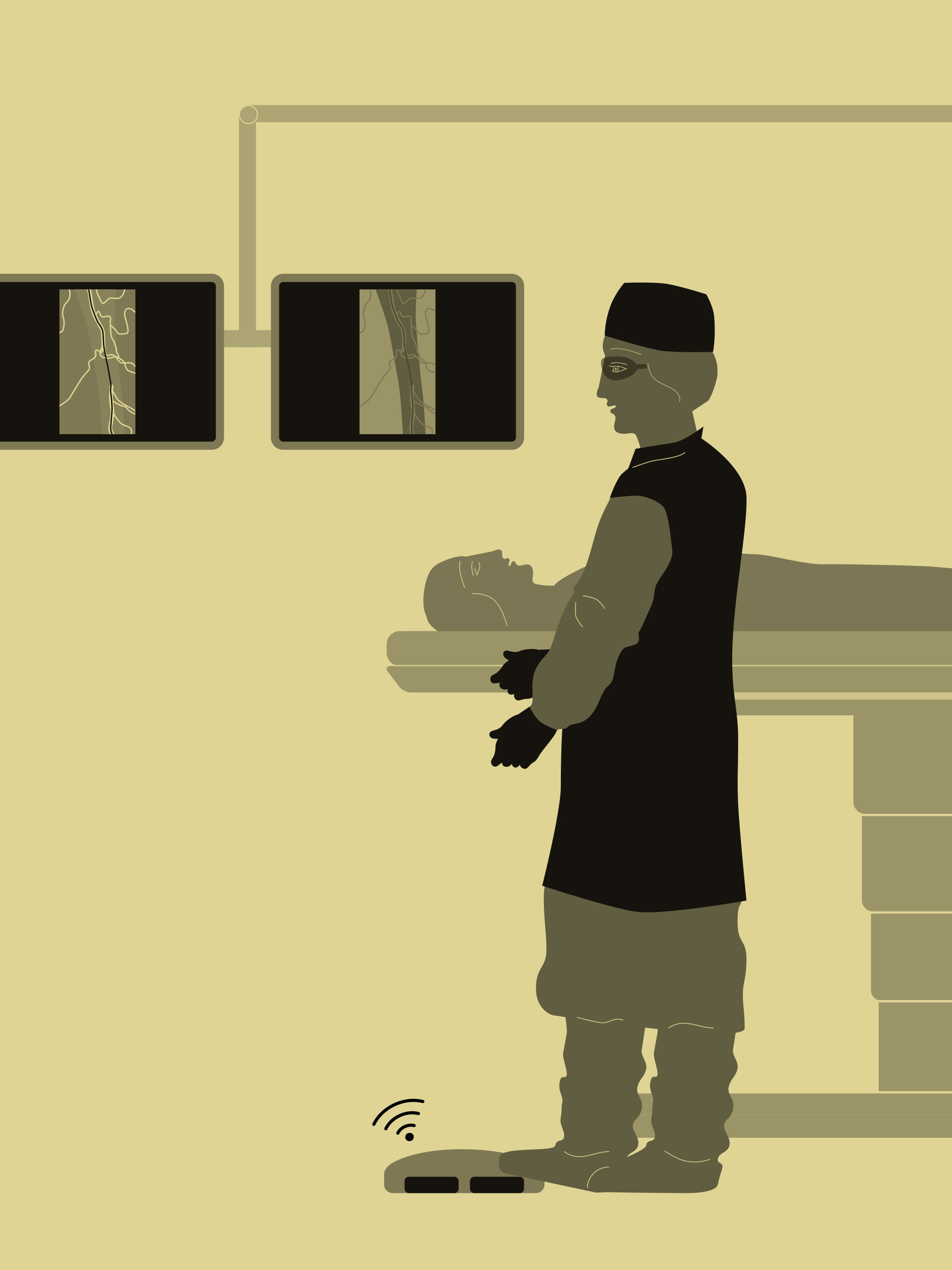
The team checks the guidewire position with a live fluoroscopic image that is merged with the panorama. The vessel roadmap is updated in real time. If the table or the patient moves, the software relocalizes the live image using the 2D-2D registration. Dr. Keschenau finds this particularly helpful in her daily work: “If a patient moves a little during sedation, we can quickly anatomically reorient and continue the operation without much effort, for example, with subsequent angiographies."
Pass the lesion with the guidewire
“Overcoming the stenosis was extremely difficult in this case. The EndoNaut vessel overlay on the live image gave us the crucial information we needed to pass the lesion with the guidewire correctly.”
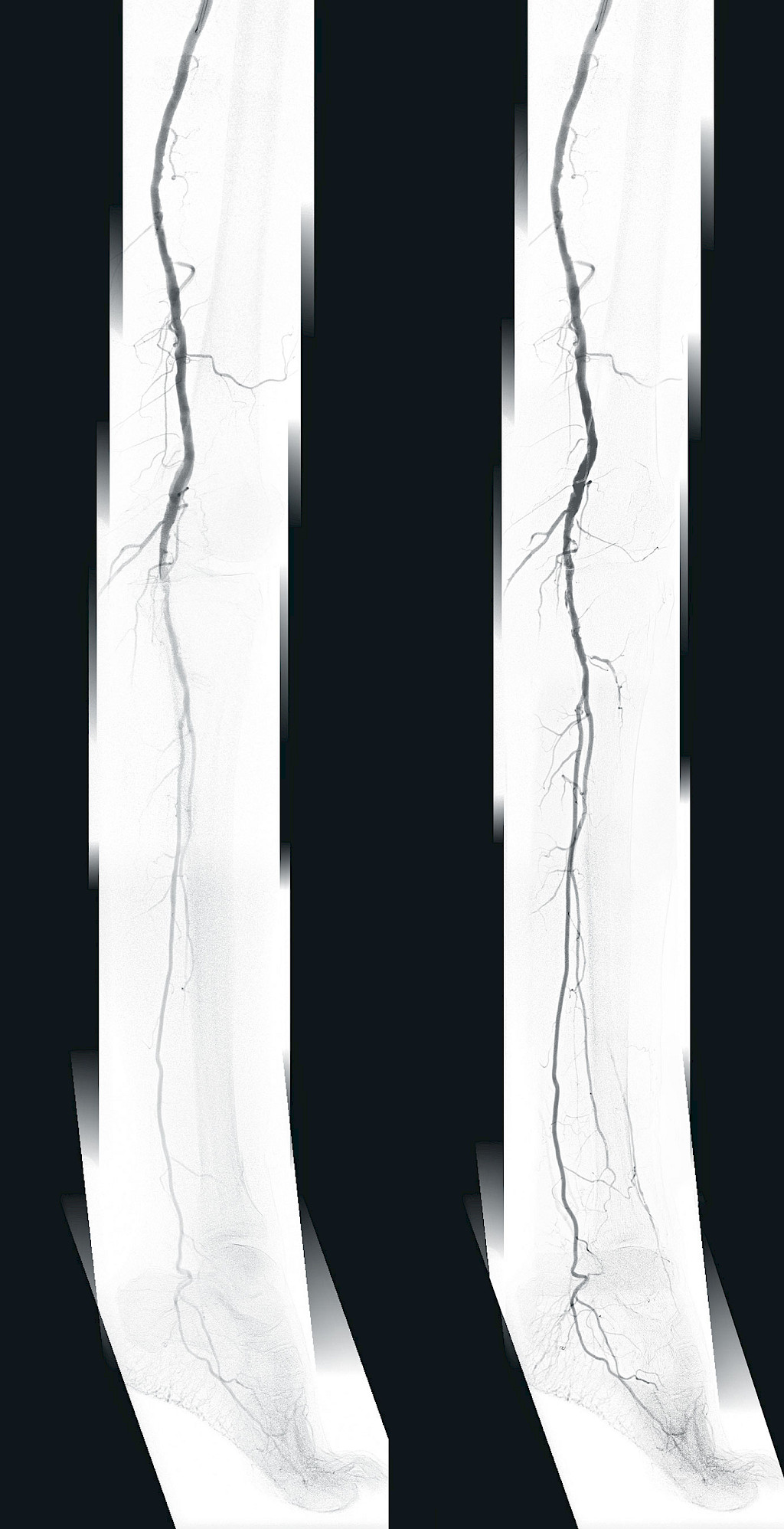
The team also makes frequent use of the workstation’s digital zoom feature. In this case, the occlusion treated with a stent during a previous procedure and the restenosis are clearly visible. Using the digital zoom, any area of the live image can be continuously enlarged. After the team has passed the stenosis, they initiate lysis to locally dissolve the vascular occlusion. After 24 hours of lysis, the team inserts a new stent.
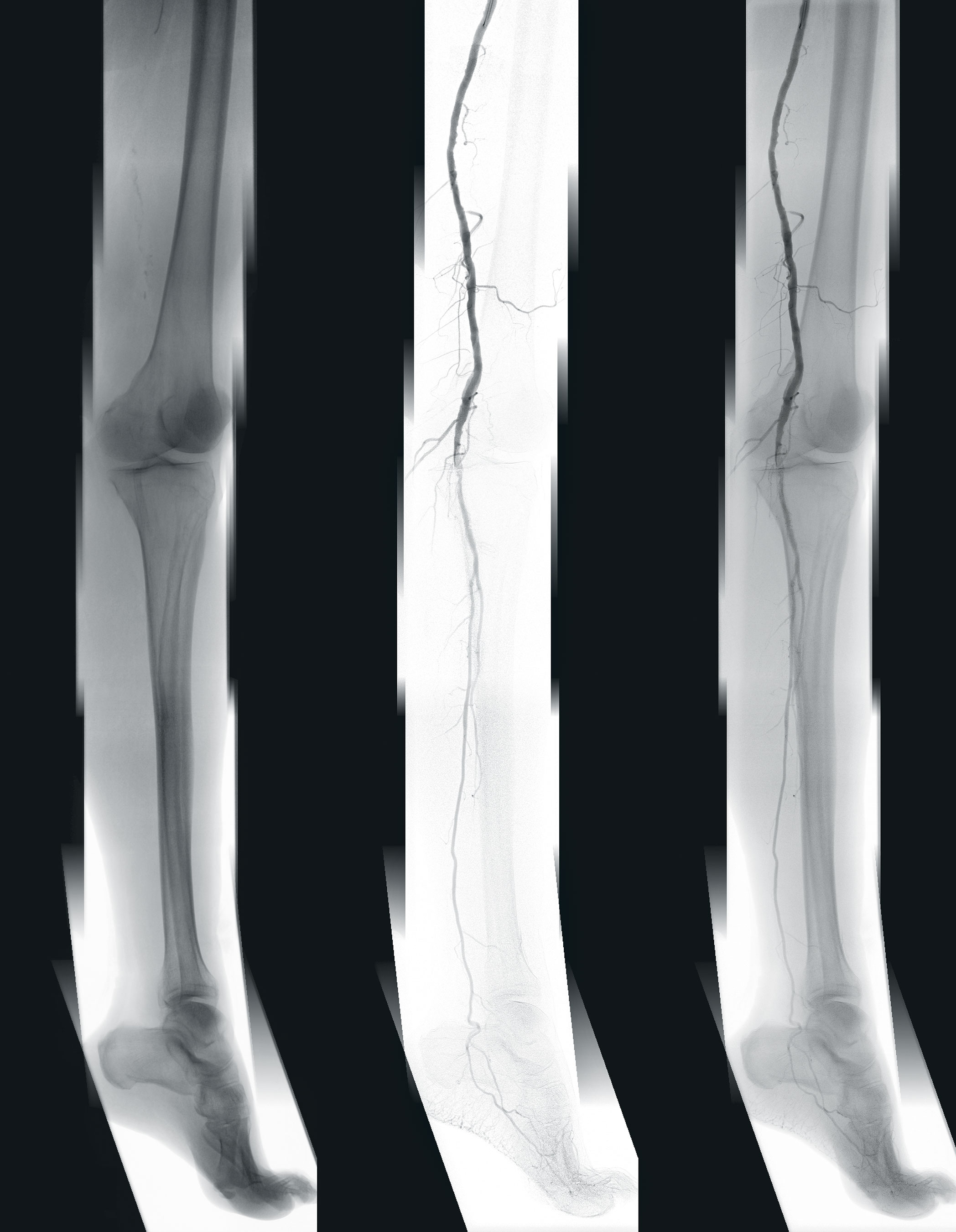
Before the first intervention, only the fibular artery was patent to mid-calf. After the procedure, the fibular artery is visible all the way to the foot, and the dorsalis pedis artery is supported by the fibular artery collaterals and well displayed in the angiography. Blood flow to the leg is restored and the patient is finally able to walk without pain, and sleep through the night.
The team is pleased with the intervention outcome. “The EndoNaut vessel overlay on the live image provided the crucial information to pass through the stenosis,” said Prof. Kalder after the procedure.
Disclaimer
1
Ziehm Vision RFD Hybrid Edition represents a group of optional hardware and software that creates an option package on the device named Ziehm Vision RFD.
2
EndoNaut® is a registered trademark of Therenva SAS. In the USA, the EndoNaut® software obtained a substantial equivalence determination and FDA clearance through the CDRH premarket notification process (510(K)). In Europe, the EndoNaut® software is CE marked (class IIb), not eligible for reimbursement. The information provided in the labelling and manual is intended for healthcare professionals only. For the safe and successful operation and use of the device, always read the instructions.