Craniomaxillofacial surgery
Highest precision even in demanding fields
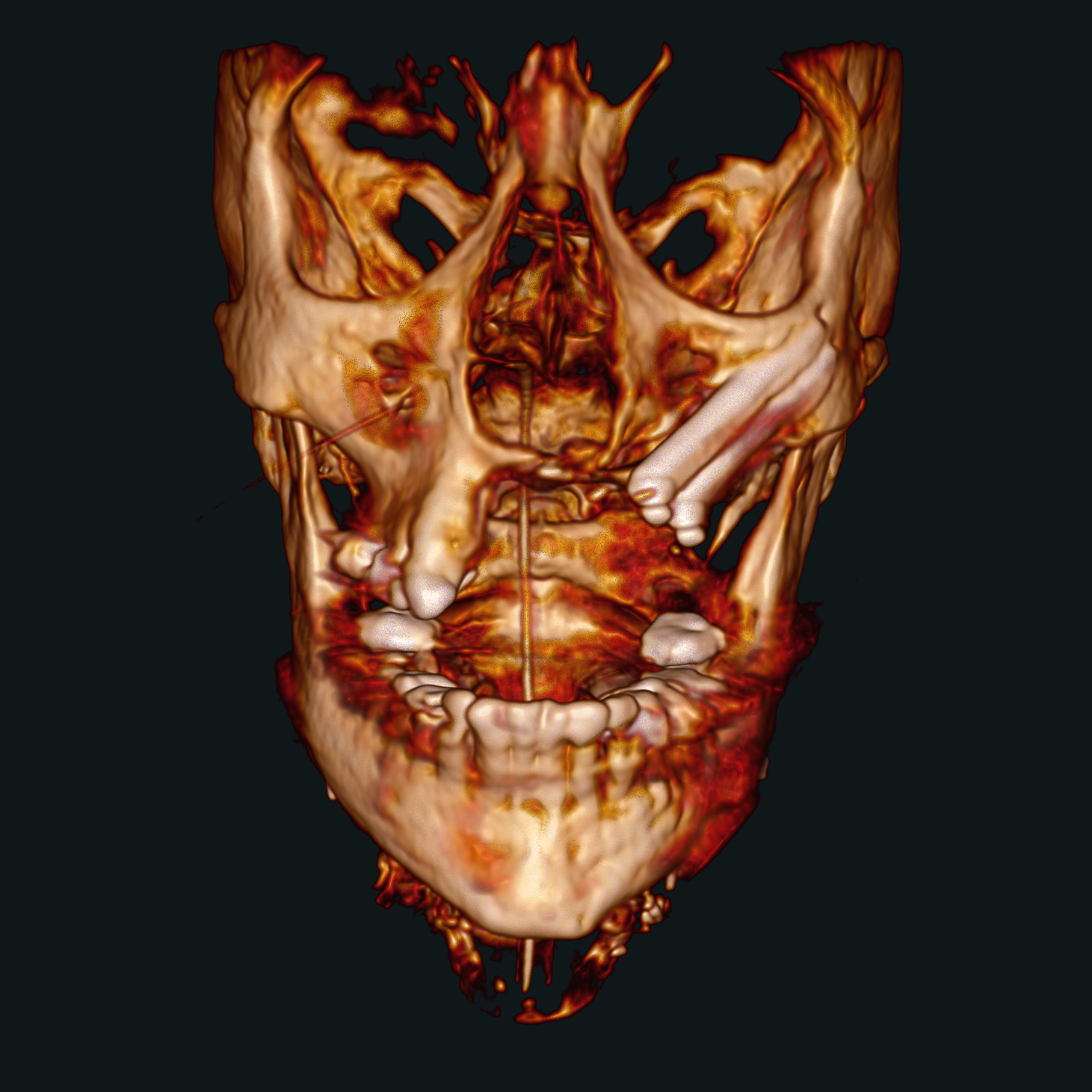
Maintaining quality control throughout all phases of surgery is vital, especially in complex cases such as midface, orbital fractures or mandibular reconstructions. Intraoperative 3D imaging with the Ziehm Vision RFD 3D enables the individual alignment of axial, coronal, and sagittal views according to the patient’s anatomy to analyze symmetry, which cannot be reliably assessed with conventional 2D imaging.
A 20 cm volume1 or a 25 cm volume2 allows for visualization of the entire skull or both orbital floors in a single 3D scan, aiding in symmetry verification of reconstructed anatomy.
Intraoperative imaging enables confirmation and adjustment of fracture reduction and implant placement and therefore provides maximum accuracy by comparing real-time data with preoperative plans.
Surface rendering combined with multiplanar reconstruction enhances clarity, particularly during minimal incisions in oral and maxillofacial surgery, where precise positioning of screws and plates is crucial.
Are you curious for more?
We are looking forward to getting your inquiry!
Disclaimer
-
1
The designation „20 cm volume“ refers to an actual reconstructed 3D volume of 19.8 cm x 19.6 cm x 18.0 cm.
-
2
The designation „25 cm volume“ refers to an actual reconstructed 3D volume of 24.9 cm x 19.6 cm x 24.9 cm